

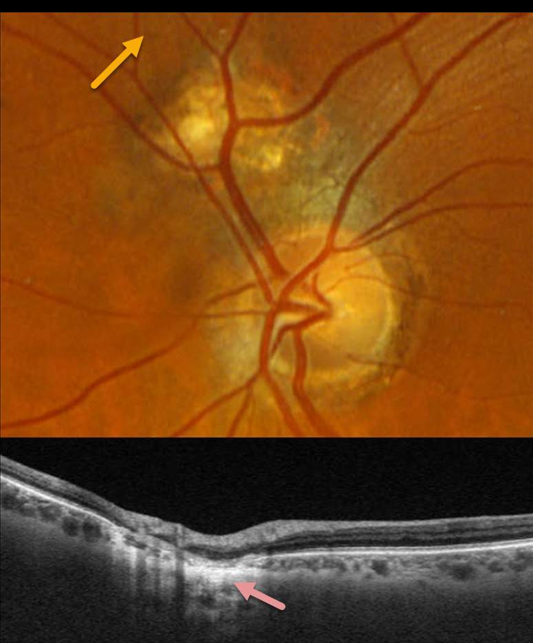
This 66YO male was referred for an asymptomatic lesion in his left fundus. Vision was 20/25 in his normal right eye and 20/25 in his left eye.
Optos color RGB imaging of the left eye shows a normal optic nerve. Just superonasal to the nerve is a round, colobomatous scar.
Triton swept-source OCT through this lesion shows disorganized neurosensory thinning with an underlying choroidal coloboma.
Learning Points:
Pseudoduplication of the optic nerve occurs when a round choroidal coloboma or chorioretinal scar about the size of the optic nerve is associated with overlying radiating blood vessels (Bloom et al, Retinal Cases 2022;16:174-176). In our experience, it is most common for toxoplasmosis scars to be located near the optic nerve.



