

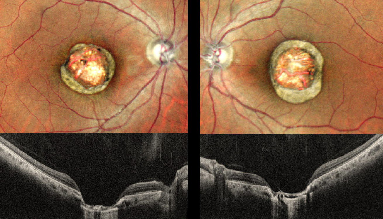
This healthy 24YO male presented with a lifelong history of stable bilateral vision loss. Vision was 20/80 OU.
Pseudocolor SLO imaging shows symmetrical macular colobomas. OCT scanning shows near-total choroidal loss within the coloboma, with a thinned, atrophic retina draped against bare sclera. Prior serologic testing elsewhere was negative for both toxoplasmosis and cytomegalovirus.
Learning Points:
Choroidal colobomas are most commonly due to failure of the optic vesicle and choroidal fissure to close during embryonic development and are usually found inferonasally. They can present as solitary or multiple lesions.
The differential diagnosis for congenital macular coloboma also includes post-inflammatory lesions (toxoplasmosis, cytomegalovirus, and Zika) and retinal dystrophies, including North Carolina macular dystrophy (NCMD). The retina within the coloboma is variably thinned, increasing the risk of rhegmatogenous retinal detachment (RRD).
The symmetry of the lesions argues for NCMD, but unfortunately, genetic testing was not performed due to cost.



