

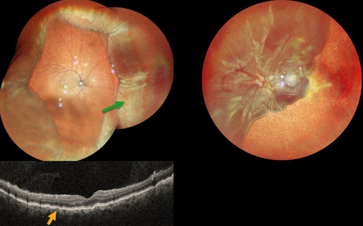
This 52YO male presented with 2 weeks of vision loss OD. He had a complicated ocular history of multiple prior surgeries elsewhere, including cataract surgery OD 2 months earlier and multiple retinal detachment surgeries OS. Vision was 20/30 OD and LP OS. Pressure was 8mmHg with no wound leaks on slit lamp OD.
MultiColor imaging shows encircling peripheral choroidal detachments OD, and OCT shows shallow macular chorioretinal folds. A total retinal detachment is noted OS with bare choroid nasally and inferiorly.
Learning Points:
A choroidal detachment is defined by the abnormal presence of fluid or blood in the suprachoroidal space. Serous choroidal detachments, also known as choroidal effusions, are a frequent complication of glaucoma surgery or following any intraocular procedure. Frank wound leaks usually require surgical repair. Otherwise, conservative management, often with topical/systemic steroids, is recommended.
Since our patient’s surgical wound was secure and the anterior chamber was formed, topical and oral steroids were prescribed.



