

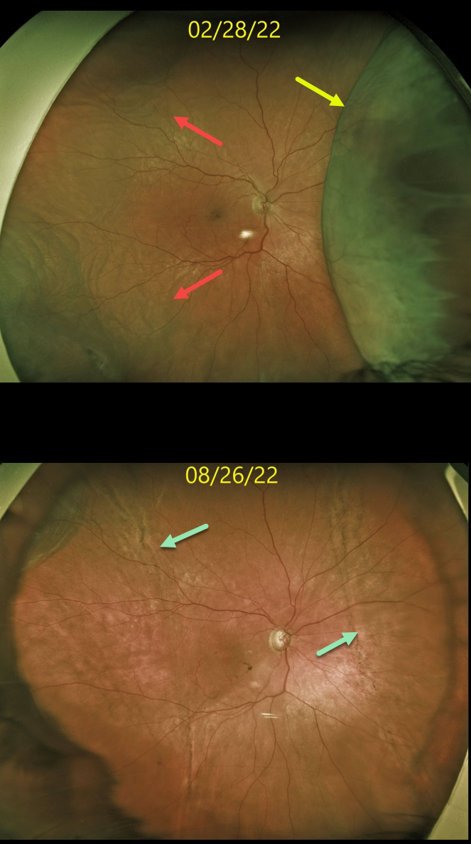
This 74YO female was receiving periocular and intravitreal steroids for persistent pseudophakic cystoid macular edema (CME). Steroid response prevented further treatment, and she eventually required XenGel glaucoma surgery to control her pressure and allow for continued CME treatment.
Four days following glaucoma surgery, vision was 20/200, and the intraocular pressure (IOP) was 6 mmHg. Optos imaging shows a large nasal choroidal detachment with a shallow temporal choroidal detachment.
Over the next 3 months, her intraocular pressure normalized with total resolution of the choroidals. A ‘high-water’ mark of subretinal pigmentation is seen along the posterior extent of both the prior nasal and temporal choroidal detachments.
Learning Points:
Choroidal detachment is relatively common immediately following glaucoma surgery.
Two mechanisms are believed to be at play: hypotony allows fluid to accumulate in the suprachoroidal space, and inflammation increases the choroidal permeability. The detached ciliary body may also produce less aqueous humor, thus creating a self-perpetuating cycle.
Often, observation is sufficient, but topical steroids and cycloplegics can be helpful in promoting resolution.



