

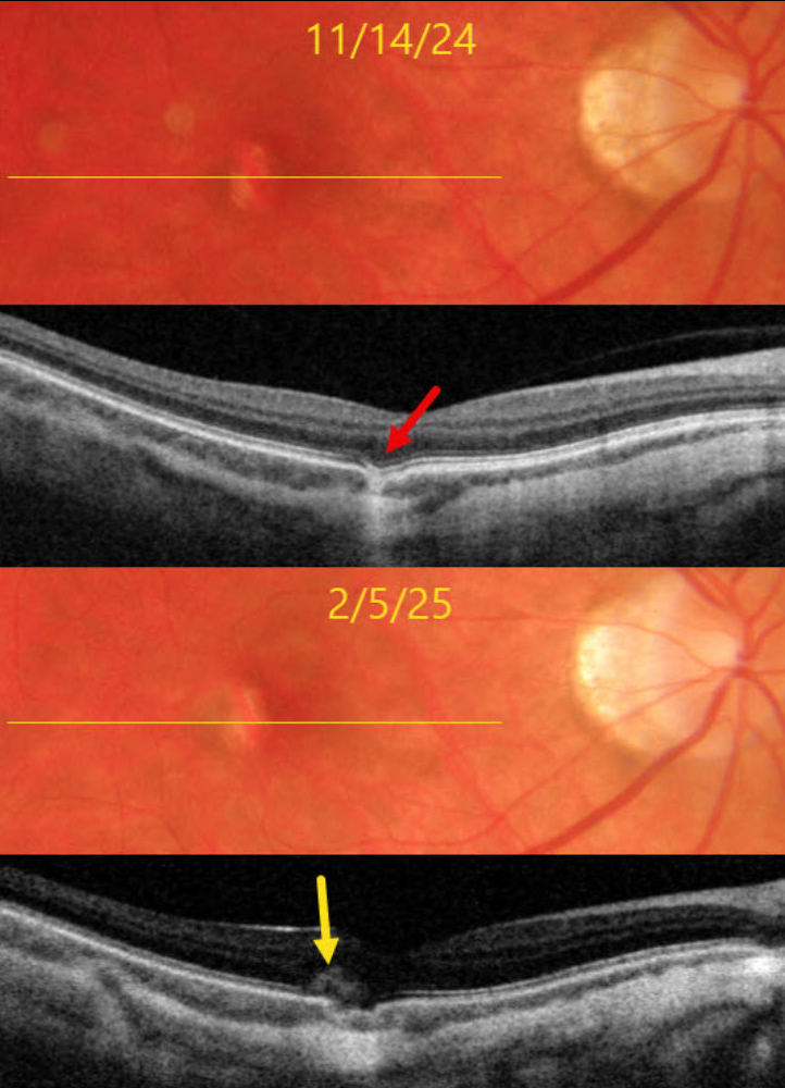
This 39YO myopic female was referred on 11/14/24 for an asymptomatic lesion in her right macula. Vision was 20/25 OD and 20/25 in her normal OS.
Triton color imaging shows a small vertically oval area of subretinal pigment loss temporal to the right fovea. Swept-source OCT shows a focal choroidal excavation (FCE) within this region.
She returned on 2/5/25 with recent blurred vision in this eye. Vision was 20/30. A new type 2 (above the RPE) macular neovascularization (MNV) is noted along the temporal edge of the FCE.
Learning Points:
FCE, originally described by Jampol et al. in 2006, is likely part of the pachychoroid spectrum, as it is usually associated with a thickened choroid and pachyvessels (not noted in our myopic patient). The FCE can be observed, although symptomatic manifestations, including active central serous retinopathy or MNV (as in our patient), often require treatment. An excellent review is by Verma et al, Br J Ophthalmol 2021;105:1043-1048.
One month after a single intravitreal Avastin injection, vision was 20/30, her symptoms had resolved, and the lesion was completely dry on funduscopy and OCT. We are following her closely with PRN therapy.



