

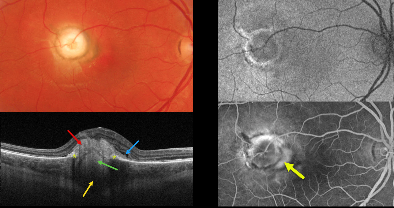
This healthy 26YO male presented with a 4-month history of decreased vision in his right eye. Vision was 20/200 OD and 20/25 in his normal OS.
Optos color RG imaging shows a well-defined, 1800-micron, pale, subretinal nodular lesion with surrounding pigment. Triton swept-source OCT shows a hyperreflective subretinal mound, a hyperreflective lesion extending from the choroid through a break in the RPE, posterior choroidal thickening and shadowing, and trace outer retinal fluid.
Optos fundus autofluorescence (FAF) shows the lesion to be hypo-FAF with a rim of hyper-FAF.
On fluorescein angiography, the nodule stains along its edges, with a leaking macular neovascularization along its nasal edge. Anti-VEGF therapy was begun.
Learning Points:
Choroidal granulomas usually appear as solitary, creamy white, round subretinal lesions. They are generally hyporeflective on OCT imaging (Invernizzi et al, Retina 2015;35:525-531) and can extend into the subretinal space, as in our patient. Causes include sarcoidosis, tuberculosis, and VKH. Testing results for our patient were completely negative, including sarcoidosis, tuberculosis, Bartonella, and syphilis.



