

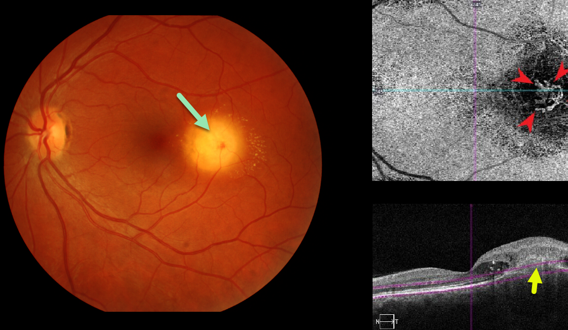
Our patient was seen recently with asymptomatic macular neovascularization (MNV) associated with a choroidal granuloma. Clinically there was new blood, fluid and lipid.
OCT angiography confirms an active MNV. This lesion is deep to the RPE which is consistent with a focal scleral nodule.
Learning Points:
Choroidal granulomas usually appear as solitary creamy white, round subretinal lesions. They are hyporeflective on OCT imaging (Invernizzi et al, Retina 2015;35:525-531). Causes include sarcoidosis, tuberculosis and VKH. They can occasionally cause exudation that responds to anti-VEGF injections or photodynamic therapy.



