

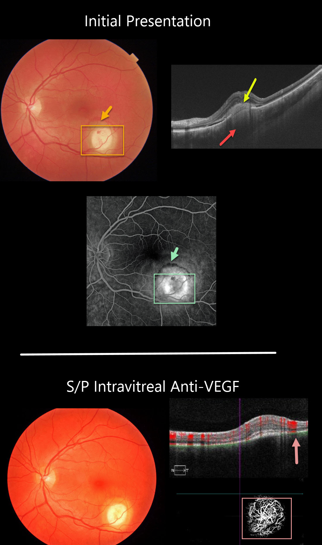
The initial photo in this series shows a focal yellow-white nodular lesion with adjacent subretinal blood. The OCT shows subretinal hyperreflective material, some shallow subretinal fluid, and an amelanotic choroidal granuloma.
Fluorescein angiography shows staining of the lesion, with some blood blockage along its superior edge.
A later fundus photo shows complete resolution of the hemorrhage following treatment with a single intravitreal anti-VEGF injection.
Despite the resolved exudation, OCTA angiography still shows flow signals within the lesion. We continue to follow her closely for recurrent exudation.
Learning Points:
Choroidal granulomas usually appear as solitary, creamy white, round subretinal lesions. They are hyporeflective on OCT imaging (Invernizzi et al, Retina 2015;35:525-531). Causes include sarcoidosis, tuberculosis and VKH. They can occasionally cause exudation that responds to anti-VEGF injections or photodynamic therapy.



