

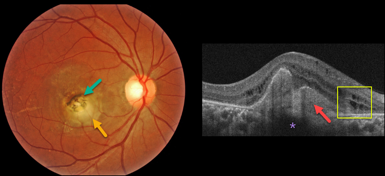
A healthy 32YO female presented with a 6-month history of vision loss in her right eye. Vision was counting fingers OD and 20/20 in her healthy left eye.
Fundus imaging shows a slightly elevated, creamy white, subfoveal lesion surrounded by slightly hyperpigmented macular scarring.
OCT shows a hyperreflective subretinal lesion that shadows the underlying choroid. There are some mild cystic changes in the inner and outer nuclear layers.
Learning Points:
Choroidal granulomas usually appear as solitary, creamy white, round subretinal lesions. They are hyporeflective on OCT imaging (Invernizzi et al, Retina 2015;35:525-531). They can extend into the subretinal space, as in our patient. Causes include sarcoidosis, tuberculosis, and VKH. Testing results for sarcoidosis and tuberculosis are still pending.



