

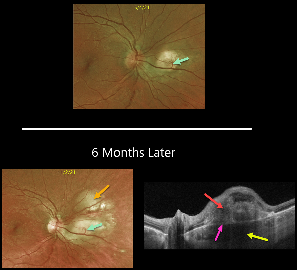
This is a follow-up from our 6/7/21 post of a choroidal granuloma, likely representing the initial manifestation of systemic sarcoidosis. The patient was sent to pulmonology to confirm the diagnosis, but he did not keep the appointment.
Six months later, the choroidal granuloma had significantly enlarged.
A retinochoroidal anastomosis is noted between the overlying retinal vein and the lesion. Triton swept-source OCT shows a thickened, hyporeflective choroidal lesion, a hyperreflective outer retinal lesion with overlying inner retinal disorganization, and visible Bruch’s membrane separating the choroidal and retinal components.
Learning Points:
Choroidal granulomas are aggregations of inflammatory cells that appear clinically as a solitary, creamy-white, round subretinal lesion. Causes include sarcoidosis, tuberculosis, and VKH.
They are hyporeflective with increased transmission on OCT imaging (see Invernizzi et al, Retina 2015;35:525-531).



