

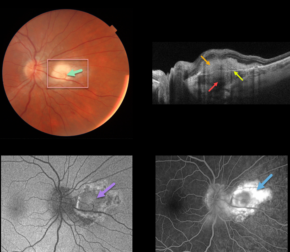
This 30YO male was referred for an asymptomatic lesion in his right eye. Vision was 20/20 OU, and the left fundus was healthy.
There is a nodular, creamy white, deep retinal/subretinal lesion just nasal to the right optic nerve. A retinochoroidal anastomosis was noted between the overlying retinal vein and the lesion.
Triton swept-source OCT shows a thickened hyporeflective choroidal lesion, a hyperreflective outer retinal lesion with overlying inner retinal disorganization, and visible Bruch’s membrane separating the choroidal and retinal components.
Optos fundus autofluorescence (FAF) shows relatively diffuse hypo-FAF, and fluorescein angiography shows diffuse staining of the lesion.
Our patient’s bloodwork and chest x-ray were suggestive of sarcoid. Quantiferon gold, ACE, and toxoplasmosis IgG/IgM were all negative. His primary care provider was alerted to the probable diagnosis of sarcoidosis.
Learning Points:
Choroidal granulomas usually appear as solitary, creamy white, round subretinal lesions. They are hyporeflective on OCT imaging (Invernizzi et al, Retina 2015;35:525-531). Causes include sarcoidosis, tuberculosis, and VKH.
The choroidal and retinal circulations normally coexist without connecting to each other. A chorioretinal anastomosis breaks this rule when a retinal vessel directly connects with the choroid.
Clinically obvious chorioretinal anastomosis is most common in disciform lesions and toxoplasmosis scars. They are also a subclinical finding noted in 65% of eyes with MacTel 2 using OCT and OCT angiography (see Spaide et al, Retina 2018;38:1920-1929).
A laser-induced chorioretinal anastomosis was originally proposed as a possible treatment for central retinal vein occlusion in 1995, and a recent randomized trial showed it could reduce the anti-VEGF treatment burden (see McAllister et al., JAMA Ophthalmol 2018;136;1391-1397). However, it is still rarely used in current clinical practice.



