

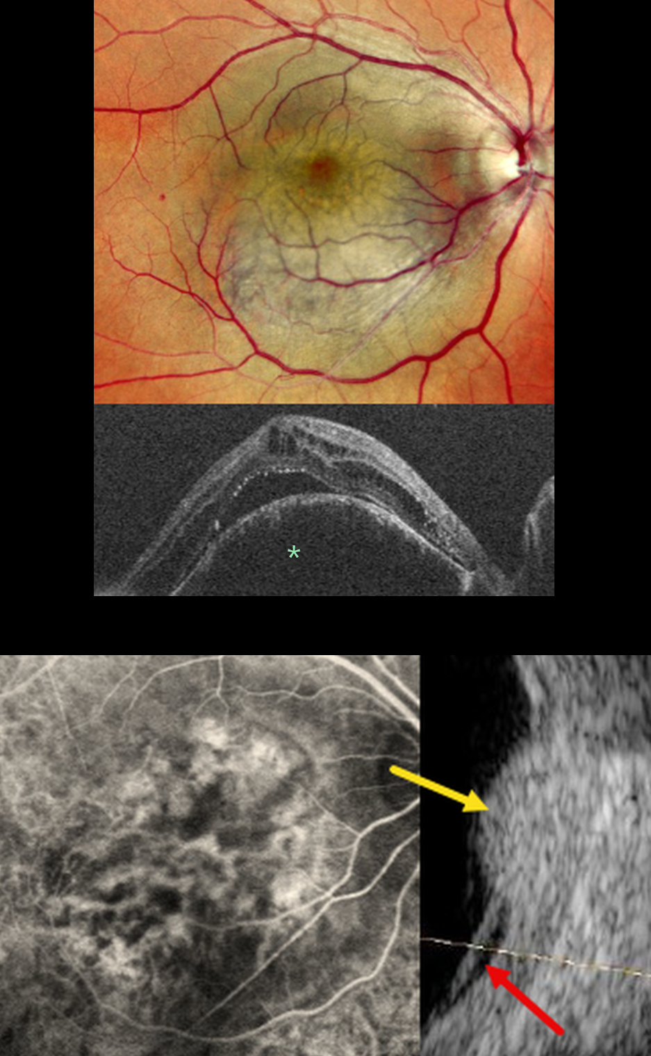
This 42YO male presented with two months of vision loss in his right eye. Vision was 20/200 OD and 20/20 in his normal left eye.
Mirante pseudocolor SLO imaging shows a pigmented subretinal lesion occupying the entire macula. OCT shows a markedly thickened and elevated choroid with overlying cystic retinal edema and subretinal fluid. The lesion is hyperfluorescent on angiography. B-scan ultrasonography shows it to have homogeneous high internal reflectivity with localized subretinal fluid inferiorly.
Learning Points:
Choroidal hemangioma is an uncommon benign hamartomatous disorder that presents as either a circumscribed, orange-red, round choroidal lesion (as in our patient), which is almost always isolated and nonsyndromic, or a diffuse form that is usually part of Sturge-Weber syndrome.
Although it has no malignant potential, it can cause vision loss due to exudation of subretinal and intraretinal fluid, which can be treated with photodynamic therapy (see Tsipursky et al., Surv Ophthalmology 2011;56:68-85).
The Mirante SLO uses blue (488nm), green (532nm), and red (670nm) confocal lasers to generate a fundus image. The blue laser best images the vitreoretinal interface and inner retina, the green images the mid retina, and the infrared images the outer retina, RPE, and choroid (Roy et al, Surv Ophthalmology 2024;69:378-402). Combining these 3 wavelengths into a single file produces a pseudocolor image that often provides the clinician with more information than a standard color photo. However, in some disorders, including choroidal hemangioma, this technology creates a false hyperpigmented appearance, which can lead to the misdiagnosis of uveal melanoma (Becker et al, OSLIR 2023;54:292-296).



