

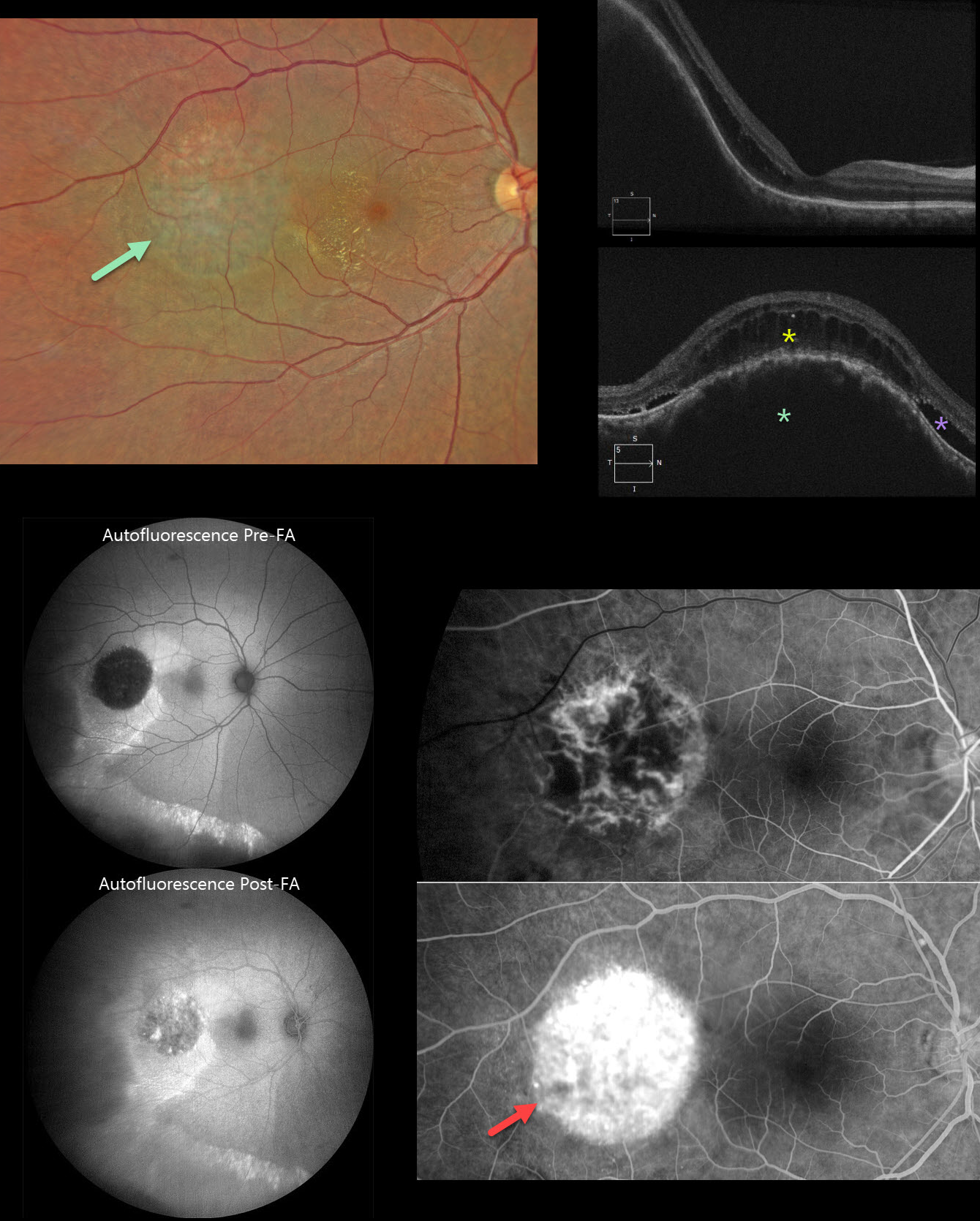
This 37YO male presented with a 3-week history of decreased vision in his right eye. Vision was 20/70 OD and 20/20 in his normal OS.
Color photography shows a subretinal lesion with a somewhat orange hue in the temporal macula. Lipid extends into the temporal fovea. Some subtle pigmentary changes extend inferiorly. OCT scanning shows marked choroidal thickening within the lesion with overlying cystic retinal edema and some subretinal fluid.
On fundus autofluorescence (FAF), the lesion is hypo-FAF with surrounding hyper-FAF and a hyper-FAF gutter extending inferiorly. The lesion becomes hyperfluorescent following fundus fluorescein angiography (FFA). The lesion shows intrinsic vasculature with late leakage on FFA. Unfortunately, our patient was lost to follow-up immediately.
Learning Points:
Choroidal hemangioma is an uncommon benign hamartomatous disorder that presents as either a circumscribed, orange-red, round choroidal lesion (our patient), which is almost always isolated and nonsyndromic, or a diffuse form, which is usually part of Sturge-Weber syndrome. Although there is no malignant potential, it can cause vision loss due to exudation of subretinal and intraretinal fluid, which is best treated with photodynamic therapy (see Yang et al, Surv Ophthalmology 2025;70:389-400). Lipid exudate is a rare finding.
Our case also reminds us why fundus imaging is performed in a specific order: color photography, followed by FAF, then FFA. In FAF, lipofuscin within the RPE is the intrinsic pigment that generates the FAF signal. In FFA, fluorescence is generated by sodium fluorescein that is injected intravenously. Our patient’s hemangioma is hypo-FAF, but when fluorescein stains the lesion, it no longer generates an ‘autofluorescent’ signal due to the fluorescein dye.



