

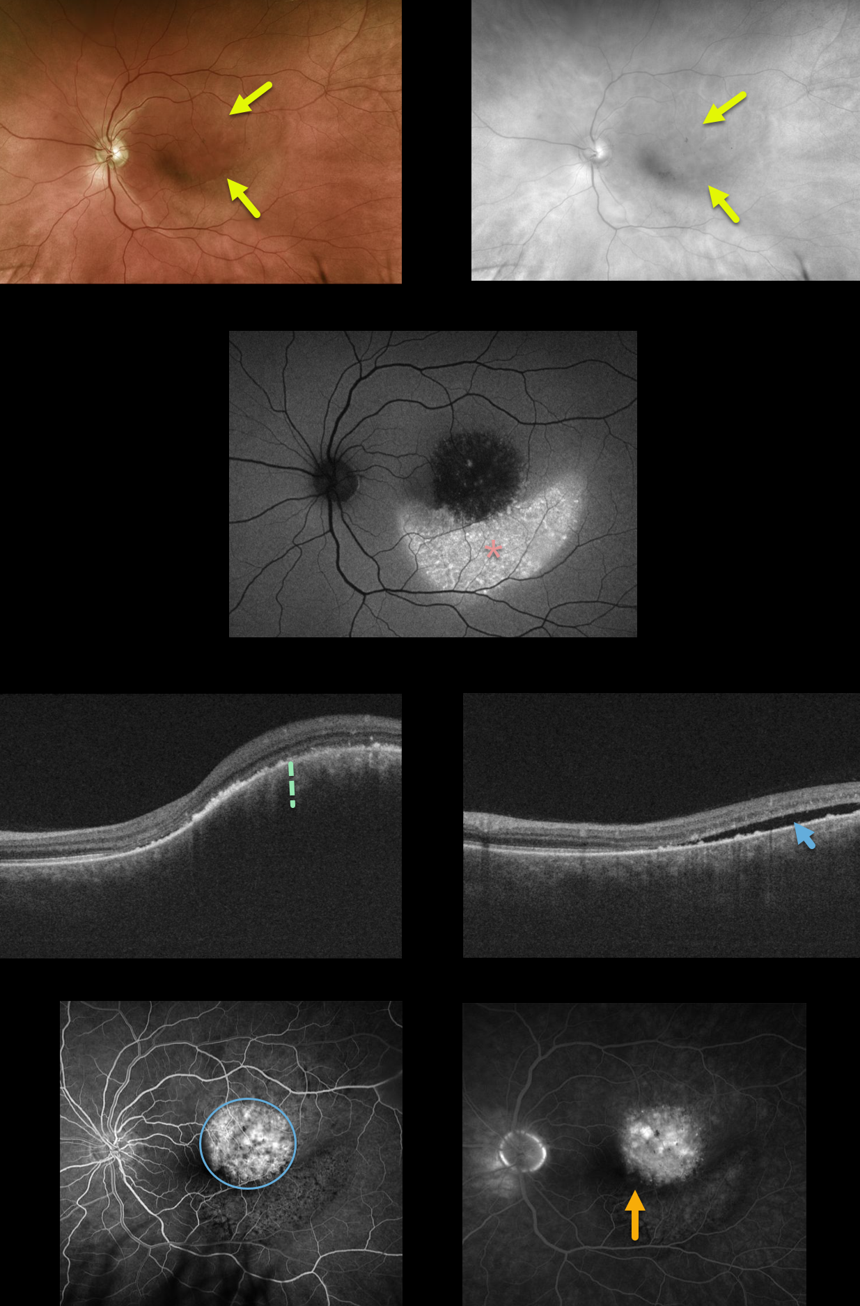
Our patient has a choroidal hemangioma which is difficult to see in the Optos color photograph, but somewhat better defined in the red channel image.
There is marked increased fundus autofluorescence inferior to the tumor, due to migrating subretinal fluid causing RPE alterations. OCT shows marked choroidal thickening within the tumor with subretinal fluid.
Fluoroscein angiography shows mild leakage from the tumor, with faint pigmentary changes inferiorly. Vision is 20/40 and he is minimally symptomatic due to the lack of foveal fluid. We are currently carefully observing this lesion and will consider photodynamic therapy if increased fluid and vision loss develop.
Learning Points:
Choroidal hemangiomas are benign choroidal vascular tumors that can become symptomatic later in life from overlying exudation. The usually round tumors are variably elevated and are often difficult to see clinically since their orange color blends in with the normal surrounding fundus.



