

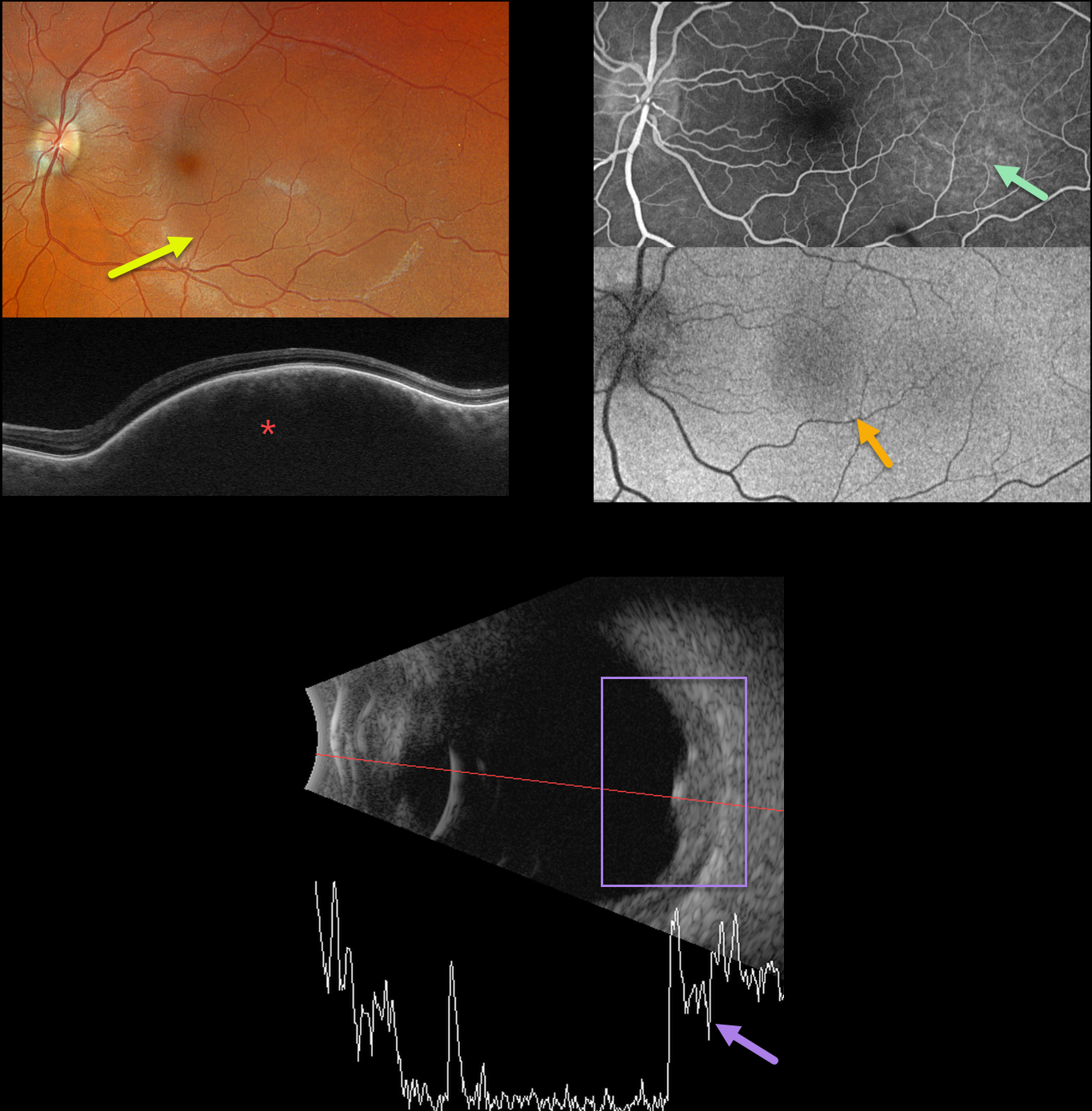
This 35YO female was referred for an asymptomatic left macular lesion. Vision was 20/20 in her normal right eye and 20/30 in her left eye.
Optos color RGB imaging shows a perfectly round subretinal lesion centered in the temporal macula, extending just beneath the macular center.
Swept-source OCT shows a thickened choroidal lesion without overlying subretinal fluid. Fluorescein angiography shows filling of this lesion without leakage, and fundus autofluorescence (FAF) shows faint hypo-FAF.
B-scan ultrasonography shows homogeneous high internal reflectivity.
Learning Points:
Isolated choroidal hemangiomas are benign vascular lesions. Although they have no malignant potential, they can cause vision loss due to exudation of subretinal and intraretinal fluid, which can be treated with photodynamic therapy (see Tsipursky et al, Surv Ophthalmology 2011;56:68-85).
Since our patient’s lesion was asymptomatic and without exudation, we will follow her several times yearly.



