

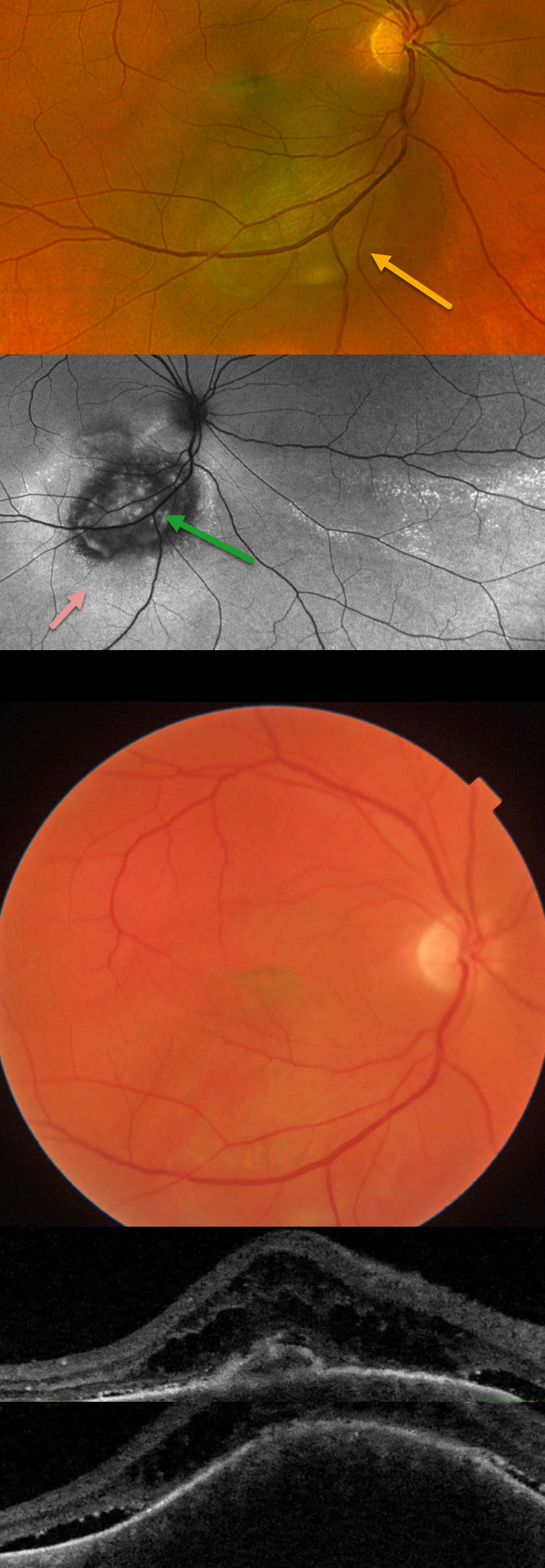
This 44YO male was referred for recent vision loss from a possible uveal melanoma. Vision was 20/150 in his right eye and 20/20 in his healthy left eye.
Optos color RG imaging shows an elevated, pigmented subretinal lesion extending from the inferior macula below the inferotemporal arcade. Fundus autofluorescence (FAF) shows this lesion to be hypo-FAF, with surrounding hyper-FAF and areas of hyper-FAF extending into the inferior and inferotemporal midperiphery, indicating prior extramacular subretinal fluid.
Triton color imaging, however, shows the choroidal lesion as orange. Swept-source OCT shows a thickened choroidal lesion with overlying subretinal fluid and cystic retinal edema. The lesion measured 9.6 x 9.0mm in basal diameter and 3.4mm thick with B-scan ultrasonography (not shown). There was moderate-to-high internal reflectivity, consistent with a choroidal hemangioma. Photodynamic therapy was recommended.
Learning Points:
Isolated choroidal hemangiomas are benign vascular lesions. Although they have no malignant potential, they can cause vision loss due to exudation of subretinal and intraretinal fluid, which can be treated with photodynamic therapy (see Tsipursky et al, Surv Ophthalmology 2011;56:68-85).
This lesion was initially misdiagnosed as a uveal malignant melanoma based on the hyperpigmented appearance on Optos RG imaging. This has also been reported in the literature (Becker et al, OSLIR 2023;54:292-296). These images are generated from a red (635nm) and green (532nm) laser. This creates a greenish tint, which distorts the true fundus color. In our patient, this was quite dramatic, with the hemangioma’s true orange color evident only in the Triton image. A newer Optos true-color RGB unit has recently been introduced, which maintains the ability to view the separate RG channels while also generating a true-to-life color image.



