

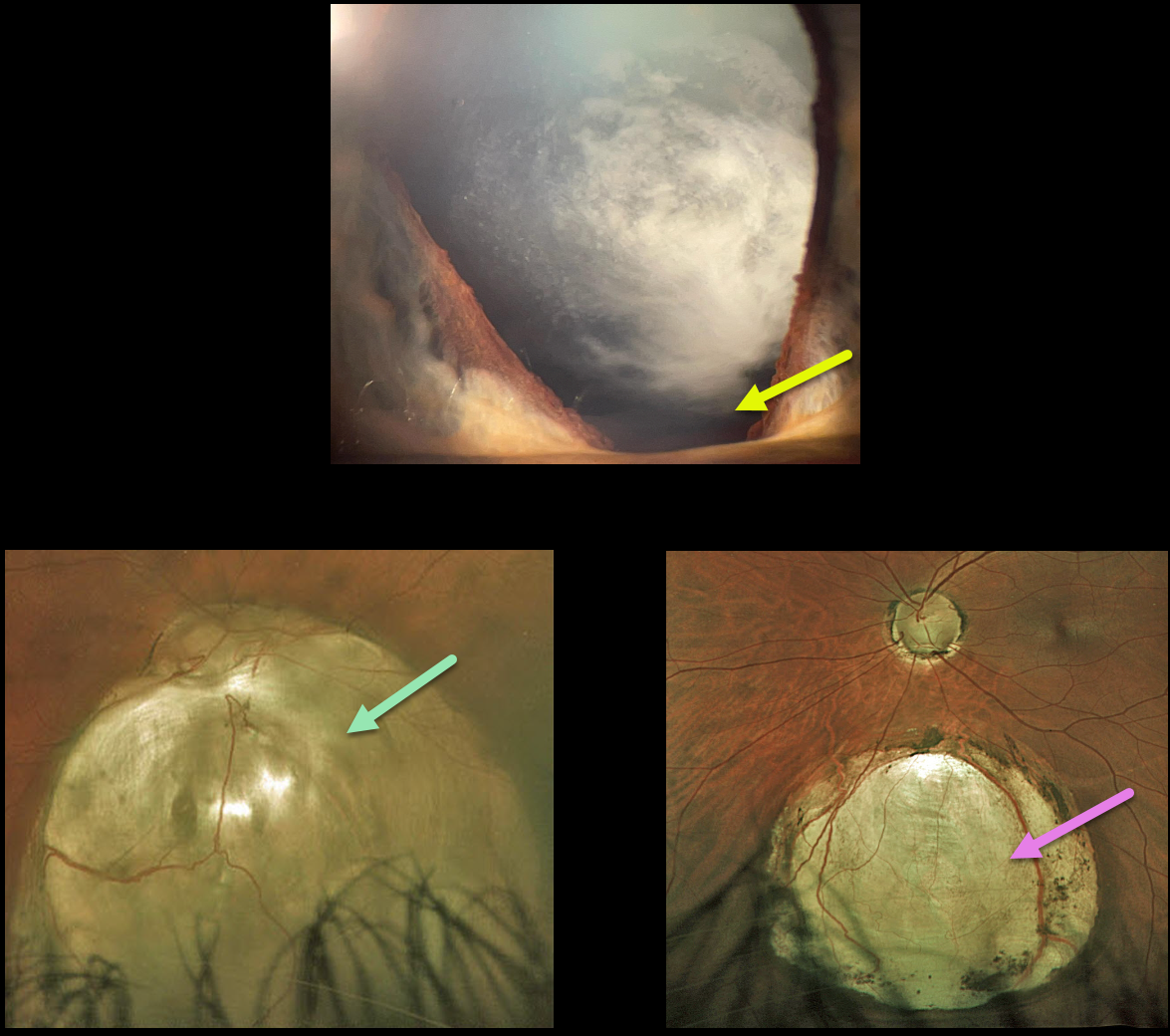
This 42YO state trooper presented with a few months of vision loss in his right eye, including significant glare with headlights. He reported a history of excellent vision throughout his life. Vision was 20/200 OD and 20/20 OS.
He had 3+ nuclear/cortical spoke cataracts consistent with this vision loss. Slit lamp examination shows an inferior iris coloboma OD, and it was normal OS.
Optos imaging of his right eye shows an inferior choroidal coloboma extending from the nerve inferiorly. The macula is normal, and OCT through the macular center is also normal (not shown). A smaller inferior midperipheral choroidal coloboma is noted in his left eye.
Cataract surgery is scheduled, and we expect his vision to return to normal, given his prior history and normal macular anatomy.
Learning Points:
Choroidal colobomas are most commonly due to failure of the optic vesicle and choroidal fissure to close during fetal development. Lesions like this may also be from other causes, including toxoplasmosis. Choroidal coloboma can present as solitary or multiple lesions. Visual prognosis depends on the location and involvement of the optic nerve and macula.



