

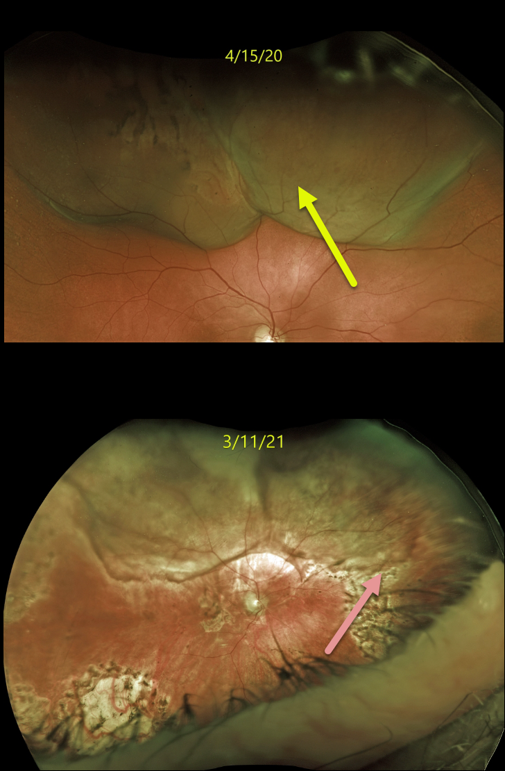
This 82YO male with a history of retinal detachment treated with a segmental buckle and cryotherapy presented on 4/15/20 with some pain and new floaters in his right eye. Vision was 20/40.
Optos imaging shows an exudative retinal detachment secondary to a large, elevated, irregular, and amelanotic choroidal mass in the superior hemisphere. B-scan ultrasonography (not shown) revealed the tumor to be 6mm in height and 23 by 13 mm in basal diameter.
There was a prior history of metastatic clear cell renal carcinoma that was treated with radical left nephrectomy and chemotherapy from 2012 to 2014. He remained in remission until 12/2019, when a thoracic lymph node was treated with external beam radiation. This new large choroidal metastatic lesion heralded more widespread metastatic disease.
A fine needle aspiration biopsy was performed, and metastatic renal cell carcinoma was confirmed. The right globe was treated with external beam radiation, and he was started on systemic immunotherapy.
After the lesion showed regression and stability, vitrectomy with silicone oil was performed for retinal detachment. When last examined on 3/11/21, the tumor had regressed. Vision was counting fingers.
Unfortunately, he died shortly after this last exam.
Learning Points:
Although uveal melanoma is the most common primary intraocular malignancy, metastatic choroidal tumors are the most common intraocular malignancy. Lung and breast cancer are the most common sources. Renal cell cancer is an uncommon cause for choroidal metastasis.



