

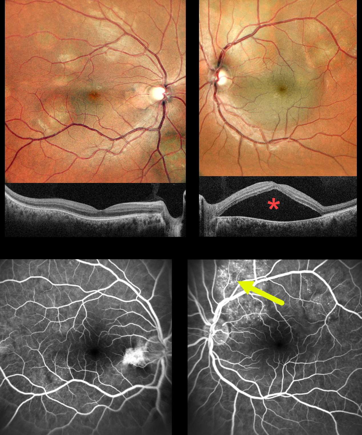
This previously healthy 30YO female presented with several weeks of decreased vision in her left eye. Vision was 20/20 OD and 20/60 OS.
MultiColor imaging shows bilateral multifocal variably sized yellow-white choroidal lesions throughout each posterior pole. A large serous detachment occupies the entire left macula, which is confirmed on SD-OCT. Fluorescein angiography of the right eye shows some speckled subretinal hyperfluorescence overlying a choroidal lesion in the distal inferotemporal macula. Angiography of the left eye shows similar but more extensive hyperfluorescence overlying the superior and superotemporal choroidal lesions, likely responsible for the serous macular fluid.
An extensive medical workup revealed widely metastatic pulmonary adenocarcinoma, and she was subsequently referred to oncology. She was unfortunately lost to follow-up immediately.
Learning Points:
Although uveal melanoma is the most common primary intraocular malignancy, metastatic choroidal tumors are the most common intraocular malignancies. Lung and breast cancer are the most common sources. The extensive multifocal lesions in each of our patient’s eyes are unusual, since most patients have 1-2 choroidal lesions (Shields et al Retina 2020;40:204-213).



