

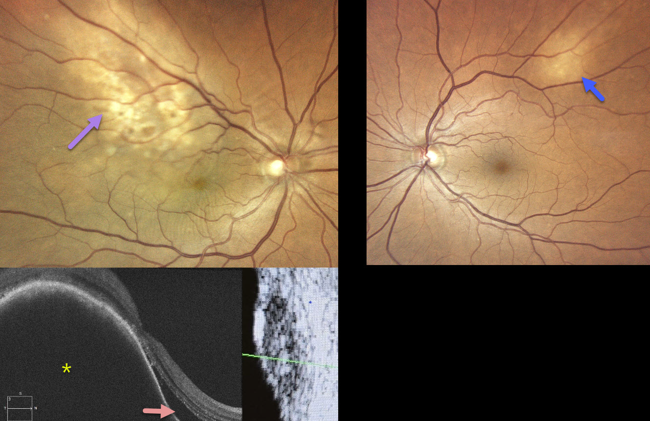
This healthy 70YO female recently underwent cataract surgery elsewhere for vision loss in her right eye. Vision failed to improve postoperatively. Vision was 20/40 OD and 20/20 OS.
Color photography OD shows an ill-defined amelanotic macular choroidal lesion extending into the superotemporal midperiphery. There are some macular chorioretinal folds. OCT scanning shows a markedly thickened choroidal lesion with overlying subretinal fluid. B-scan ultrasonography shows moderate homogenous internal reflectivity. The left eye shows a much smaller amelanotic choroidal lesion superotemporally.
MRI scanning showed a right front paramedial metastatic lesion, and she was subsequently diagnosed with a right-sided primary pulmonary neoplasia. Systemic chemotherapy with pulmonary radiotherapy began without local treatment for the choroidal metastases. When examined 5 months later, vision was no light perception OD and 20/20 OS. The right optic nerve was pale, and the metastatic lesions were fairly stable bilaterally (not shown).
Learning Points:
Although uveal melanoma is the most common primary intraocular malignancy, metastatic choroidal tumors are the most common intraocular malignancies. Lung and breast cancer are the most common sources. Although we can’t be certain, we believe that our patient’s ‘cataract’ symptoms were most likely caused by a missed choroidal metastasis. This underscores the importance of a thorough preoperative dilated funduscopic exam before cataract surgery. In our patient’s case, this not only led to an unnecessary cataract procedure but also delayed the diagnosis and treatment of her metastatic disease.



