

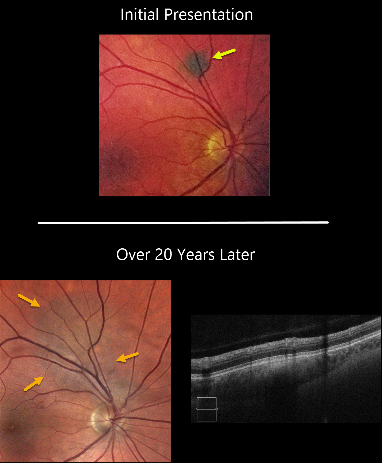
This 58YO male was initially seen in 2001 with a small asymptomatic choroidal nevus superior to the right optic nerve.
He presented to us in 2022 with a still asymptomatic but much enlarged nevus. The lesion now extends to the nerve. There is no overlying orange pigment or subretinal fluid clinically or on OCT. The lesion was 0.4mm thick.
Learning Points:
Clinical characteristics of choroidal nevi at risk for progressing to a uveal MM were initially described by Shields et al (Ophthalmology 1995;102:1351-1361). The most recent version of this famous acronym (Shields et al, Retina 2019;39:1840-1851) is To Find Small Ocular Melanoma Doing Imaging (TFSOM-DIM), which stands for:
To (Thickness >2mm)
Find (subretinal Fluid)
Small (Symptoms, vision <= 20/50)
Ocular (Orange pigment)
Melanoma (Melanoma hollow)
DIM (DIaMeter > 5mm)
Although our patient’s nevus has grown over time, this does not imply malignant transformation, as about one-third will enlarge with long-term follow-up (Shields et al., Ophthalmology 2011;118;382-388).
Shields et al found a marked increased risk for
malignant transformation for choroidal nevi over 2.0mm (Retina 2019;39:1840-1851).
Since our patient’s nevus is still well below this number and has no TFSOM-DIM risk factors other than diameter, we feel safe managing him conservatively with observation twice yearly.



