

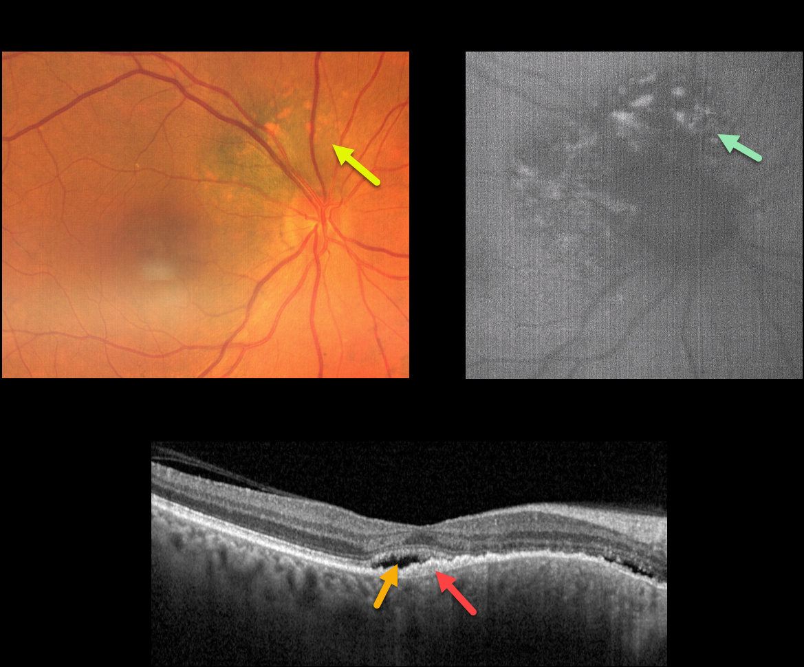
This 71YO female was referred for further treatment of chronic idiopathic central serous chorioretinopathy (ICSC) of her right eye, for which she had previously received unsuccessful anti-VEGF therapy at another institution. Vision was 20/100 OD and 20/20 in her normal left eye.
Fundus photography shows a variably pigmented, minimally thickened peripapillary choroidal nevus that extends from about 7-1 o’clock around the optic nerve margin. Overlying orange pigmentation is noted, which variably hyper-autofluoresces.
OCT scanning shows shallow foveal subretinal fluid with a subretinal layer of thickened hyperreflective material extending nasally overlying the thickened nevus. Beneath this is a very shallow, irregular elevation of the RPE.
Learning Points:
Clinical characteristics of choroidal nevi at risk of progressing to uveal malignant melanoma (MM) were initially described by Shields et al. (Ophthalmology 1995;102:1351-1361).
The most recent version of this famous acronym (Shields et al, Retina 2019;39:1840-1851) is To Find Small Ocular Melanoma Doing Imaging (TFSOM-DIM), which stands for To (Thickness >2mm) Find (subretinal Fluid) Small (Symptoms, vision <= 20/50) Ocular (Orange pigment) Melanoma (Melanoma hollow on ultrasonography) DIM (DIaMeter > 5mm).
ICSC and pachychoroid disorders, similar to syphilis, are one of the ‘great imitators,’ including uveal MM masquerading as ICSC (Negretti et al, Ophthalmology Retina 2023;7:171-177).
In our patient, on the central macular OCT B-scan, one sees a thickened choroid, subretinal fluid, and hyperreflective shed outer segments/lipofuscin, all of which are suggestive of ICSC.
This case emphasizes the importance of looking beyond the OCT, using both the clinical exam and multimodal imaging, for every patient, including those with chronic ICSC, to arrive at the correct diagnosis and treatment.
Since our patient has several high-risk characteristics (fluid, symptoms, and orange pigment), she was referred to an ocular oncologist for further management.



