

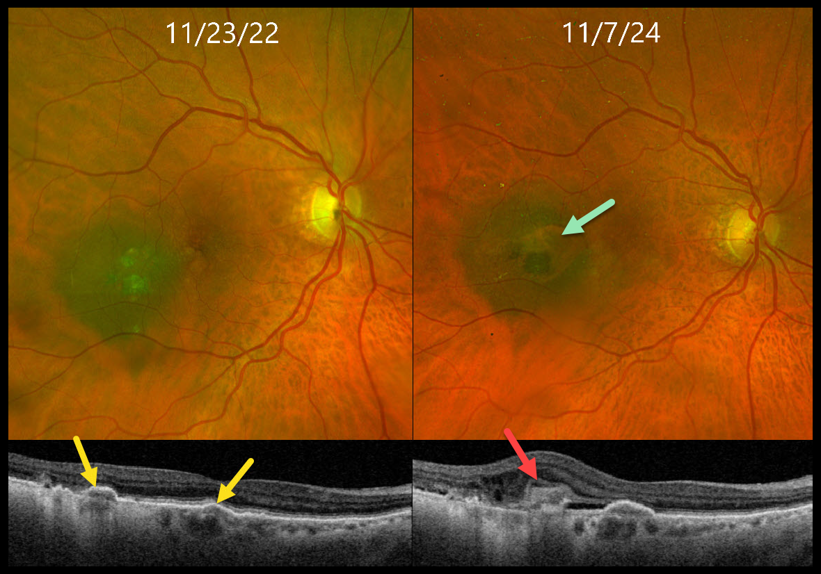
This 85YO female presented for her annual choroidal nevus examination, noticing worsening reading vision. Vision had decreased to 20/200 from 20/40 one year earlier.
When seen 2 years earlier, Triton swept-source OCT showed a few areas of drusen-like changes overlying a temporal macular nevus. At the current visit, although the margins of the nevus were unchanged, Optos color RGB imaging shows a new area of relative depigmentation with a nodular area of pigmented subretinal thickening. OCT scanning shows a hyperreflective type 2 (above the RPE) macular neovascularization (MNV) with mild associated fluid.
Monthly intravitreal Avastin therapy was started. The lesion regressed after several monthly injections, vision improved to 20/60, and a treat-and-extend regimen was started.
Learning Points:
Macular neovascularization is a rare complication of choroidal nevi. Intravitreal Avastin monotherapy is usually successful, although photodynamic therapy may be needed in some cases (Munie and Demirci, Ophthalmology Retina 2018;2:53-58).
We were initially somewhat concerned that the OCT appearance of the type 2 MNV might represent early tumor proliferation through Bruch’s membrane, but the absence of underlying choroidal thickening and the prompt response to anti-VEGF treatment argued against this.



