

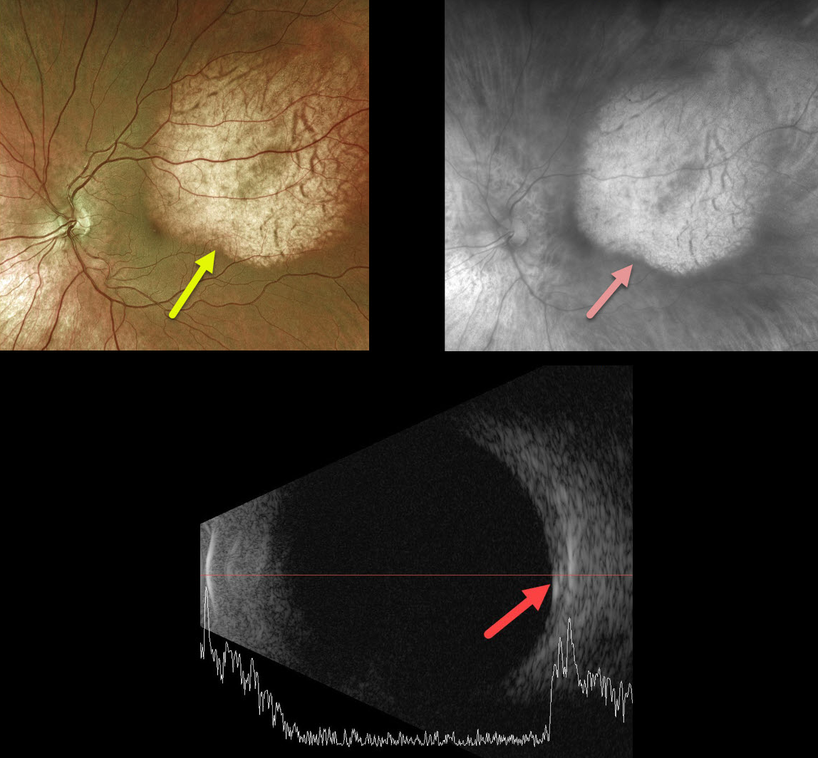
Our best guess is that this patient has a choroidal osteoma. As choroidal lesions, they are best seen on the Optos red channel.
We would, however, expect more reflectivity on the B-scan as osteomas are typically markedly hyperreflective.
Learning Points:
Choroidal osteomas are benign, calcified amelanotic choroidal tumors. They are more common in women, are almost always unilateral, and usually located in the peripapillary or macular regions.
Ultrasonography is useful in differentiating choroidal osteomas from other amelanotic choroidal tumors.
Fluorescein angiography (FA) usually shows early patchy hyperfluorescence with late diffuse staining.



