

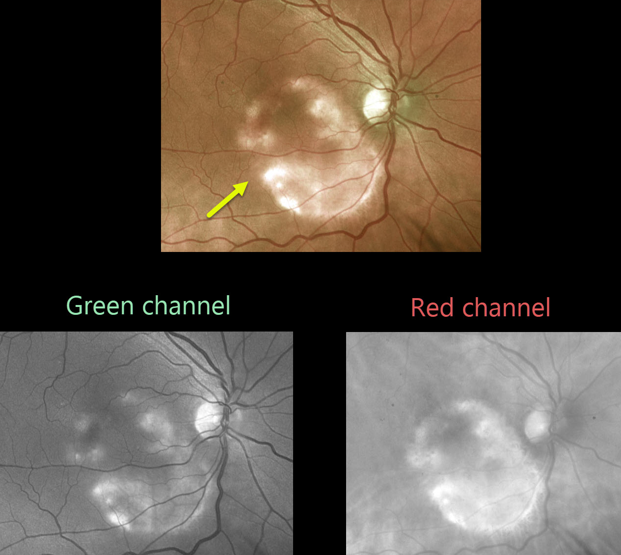
This asymptomatic patient was noted to have a choroidal osteoma.
As choroidal lesions, osteomas are best seen on the red channel compared with the green Optos channel. Ultrasonography is often helpful in differentiating an osteoma (markedly hyperreflective) from other amelanotic choroidal tumors.
Learning Points:
Choroidal osteomas are benign, calcified amelanotic choroidal tumors. They are more common in women, are almost always unilateral, and are usually located in the peripapillary or macular region.
Although many patients are asymptomatic, vision loss can occur from overlying subretinal fluid or macular neovascularization.



