

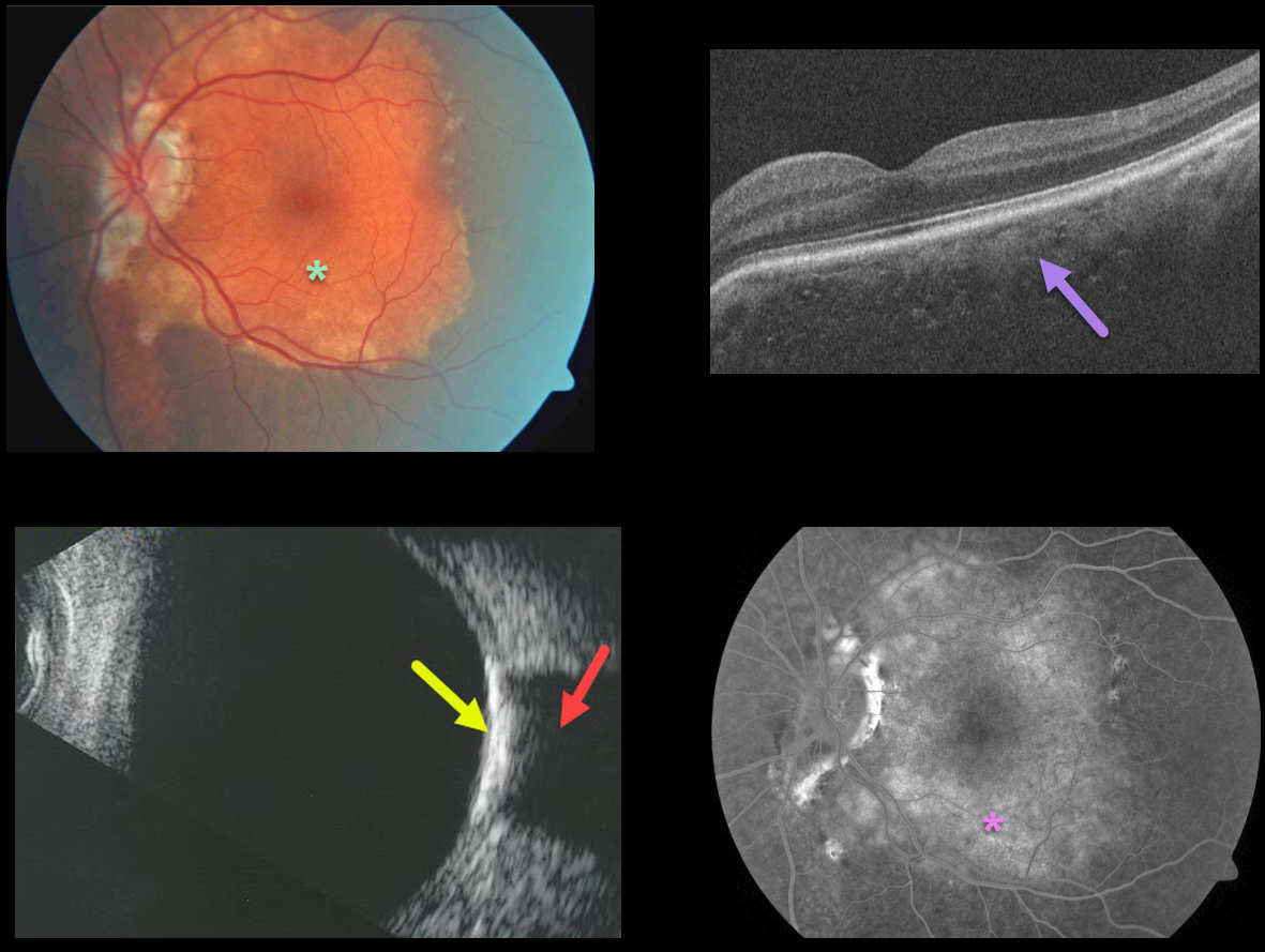
This patient presented with an orange-yellow choroidal lesion occupying the entire left macula.
OCT scanning shows diffuse choroidal thickening with small scattered specks of increased reflectivity.
B-scan ultrasonography shows marked increased reflectivity with marked posterior shadowing. The lesion diffusely stains on fluorescein angiography.
Learning Points:
Choroidal osteomas are benign, calcified amelanotic choroidal tumors. They are more common in women, are almost always unilateral, and are usually located in the peripapillary or macular regions.
Ultrasonography, as in this patient, is often helpful in differentiating an osteoma (markedly hyperreflective) from other amelanotic choroidal tumors. Fluorescein angiography usually shows early patchy hyperfluorescence with late diffuse staining.
Although the tumor itself does not require treatment, patients still need to be followed due to the risk of macular neovascularization.



