

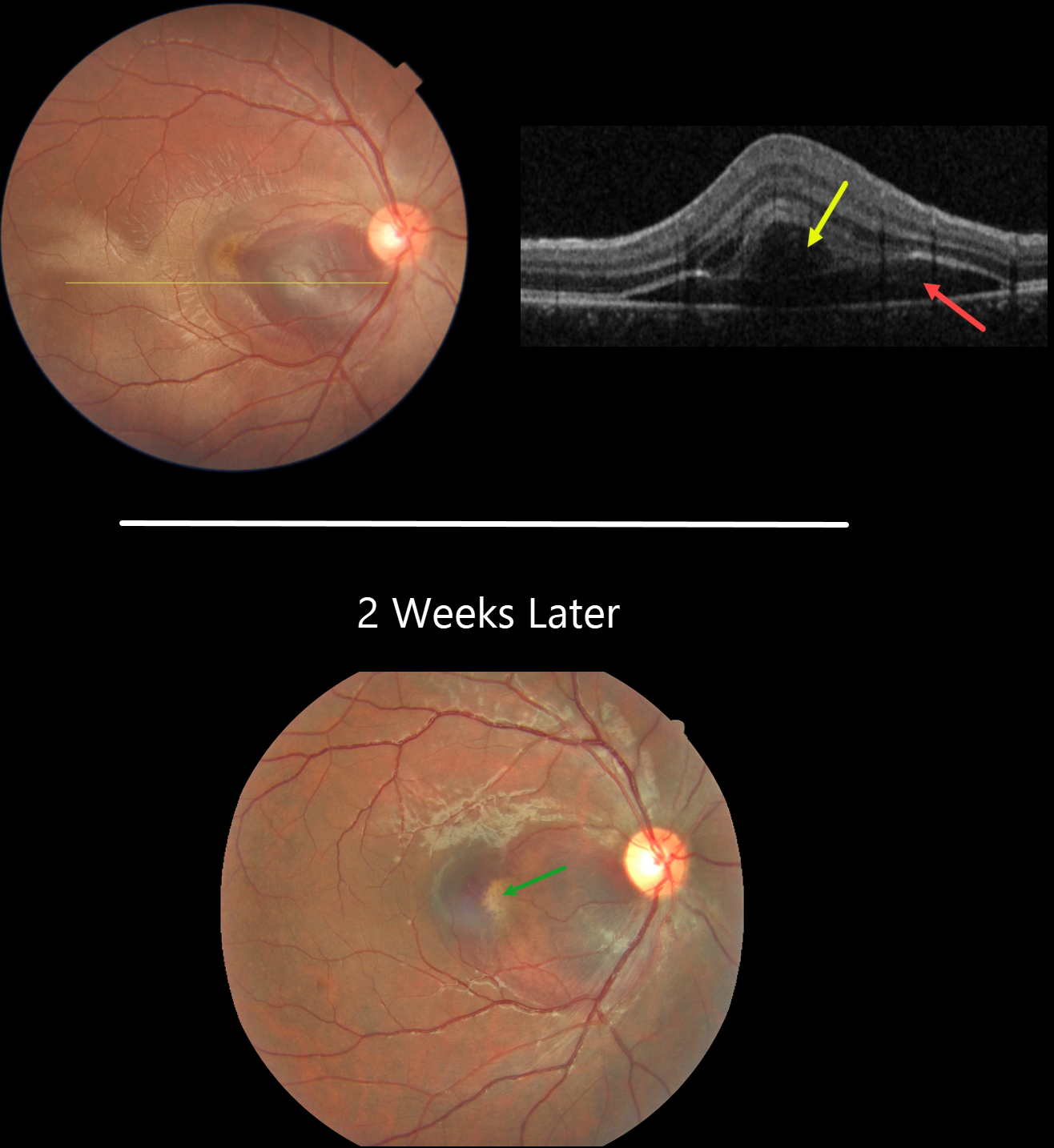
This 21YO male presented with immediate vision loss after being hit with a cricket ball in his right eye one day earlier. Vision was 20/60.
Fundus photography shows a subretinal hemorrhage in the nasal macula, a small rim of surrounding subretinal fluid, and a more temporal ill-defined commotio retina.
OCT scanning shows hyporeflective retinal elevation from the subretinal blood and fluid, along with an overlying bacillary layer detachment.
Our patient shows several findings classic for acute blunt trauma, including commotio retinae and subretinal blood. This subretinal blood almost always indicates an underlying choroidal rupture, which did, in fact, reveal itself on follow-up 2 weeks later, when vision returned to 20/20. The visual prognosis for these choroidal ruptures is good as long as the rupture spares the foveal center, although patients need lifelong surveillance due to the risk of secondary macular neovascularization.
Learning Points:
A bacillary layer (cone and rod inner and outer segments) detachment (BLD) appears as a unique dome-shaped collection of intraretinal fluid from photoreceptor splitting. These detachments can be observed in an increasing number of conditions, most classically Vogt-Koyanagi-Harada disease (Cicinelli et al, Ophthalmology Retina 2020;4:454-456).
About 8% of eyes with acute non-penetrating ocular trauma have a BLD (Venkatesh et al, Can J Ophthalmol 2022;57:328-336). These resolve within 2-10 days without visual or anatomic sequelae.



