

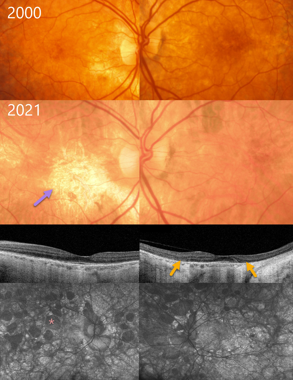
This 37YO female, whose son was diagnosed with choroideremia, was examined in 2000. Color photography shows some mild bilateral pigmentary changes.
Over the subsequent 20+ years, her ocular findings have progressed into more typical choroideremia. When last examined in 2021, vision was 20/30 OU.
The macula is now atrophic OD with minimal progressive changes OS. Swept-source OCT, however, shows fairly symmetrical outer retinal atrophy that spares each macular center. Optos autofluorescence reveals multifocal lobular areas of atrophy extending into each periphery. Genetic testing was heterozygous for a pathogenic CHM variant.
Learning Points:
Choroideremia is an X-linked recessive chorioretinal dystrophy caused by a mutation in the CHM gene located on the long arm of the X chromosome. Sons of female carriers have a 50% chance of developing choroideremia, and daughters have a 50% chance of becoming carriers. Some female carriers, as in our patient, can still develop choroideremia due to irregular inactivation of the X chromosome (see Jauregui et al, AJO 2019;207:77-86).
The onset of night blindness usually begins between the ages of 10 and 30, followed by peripheral visual field loss. Zones of patchy RPE and chorioretinal atrophy initially appear in the mid-periphery, gradually spreading anteriorly and posteriorly. Eventually, the patient is left with only a small island of vision, narrowed retinal vessels, and optic atrophy.



