

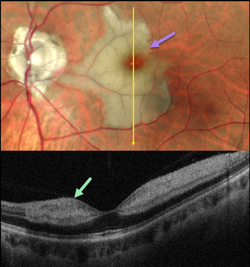
This healthy 46YO male presented with an 8-day history of a central scotoma in his left eye. Vision was 20/20 in his normal OD and 20/400 OS.
Pseudocolor SLO imaging shows an acute cilioretinal artery occlusion with foveal sparing. OCT scanning shows marked inner retinal hyperreflectivity with posterior shadowing.
Learning Points:
The inner two-thirds of the neurosensory retina is supplied by the central retinal artery, with the choroid supplying the RPE and photoreceptor layer. Cilioretinal arteries, which arise from the posterior choroidal circulation, are present in up to 50% of individuals, and bilateral in about 25% (Schneider et al, Acta Ophthalmologica 2021;99:e310-e318).
Their clinical significance usually arises from foveal preservation in eyes with concomitant retinal artery occlusions (Brown and Shields, Arch Ophthalmology 1979;97:84-92). However, they can also be a rare cause of primary branch retinal artery occlusion, either in isolation (our patient) or in association with other entities, including central retinal vein occlusions (Hayreh et al, Retina 2008;28:581-594).



