

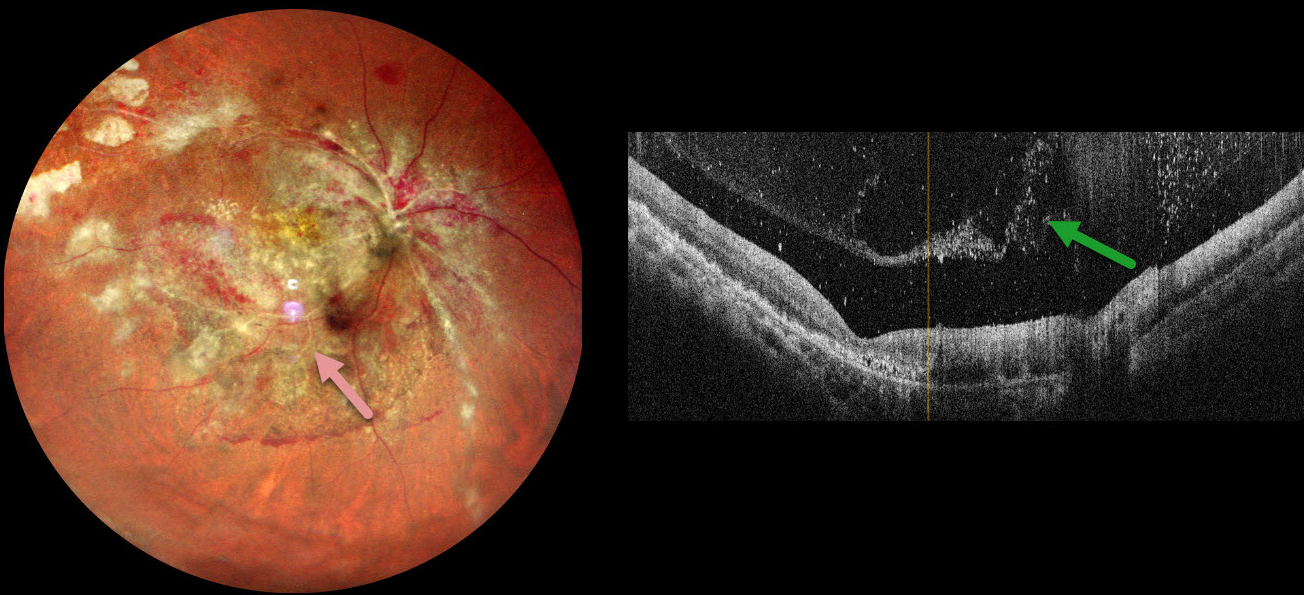
This 34YO male with a known history of HIV infection presented with 1 month of vision loss in his right eye. He was treated with a six-month course of systemic anti-tuberculous therapy for pulmonary tuberculosis two years earlier. One month earlier, he was diagnosed with HIV AIDS with a CD4 count of 43 and was started on systemic anti-retroviral therapy. Vision was counting fingers OD and 20/20 in his normal OS.
Multicolor imaging shows variably active cytomegalovirus (CMV) retinitis throughout the right posterior pole, including patches of necrotic white retina and retinal hemorrhages. The fundus is ischemic with sheathed arterioles. OCT scanning shows a vitreous separation with extensive vitreous cells.
Learning Points:
CMV retinitis develops as a reactivation of latent CMV in immunosuppressed individuals. Before effective antiretroviral treatment emerged in the mid to late 1990’s, CMV retinitis developed in up to 40% of HIV/AIDS patients, often within the last 6 months of life. Treatment includes a combination of intravenous and intravitreal medications, including ganciclovir, foscarnet, and cidofovir. Rhegmatogenous retinal detachment is a common late sequela and requires vitrectomy with silicone oil.
Our patient’s funduscopic findings are classic for CMV retinitis. However, patients with this degree of retinitis will usually have bilateral findings, and our patient’s left fundus was normal. These immunosuppressed patients also cannot mount an adequate immune response and therefore usually have a quiet vitreous, unlike our patient. It is therefore possible that there may also be an underlying tuberculous component. Unfortunately, our patient was immediately lost to follow-up before we could start intravitreal and systemic CMV therapy.



