

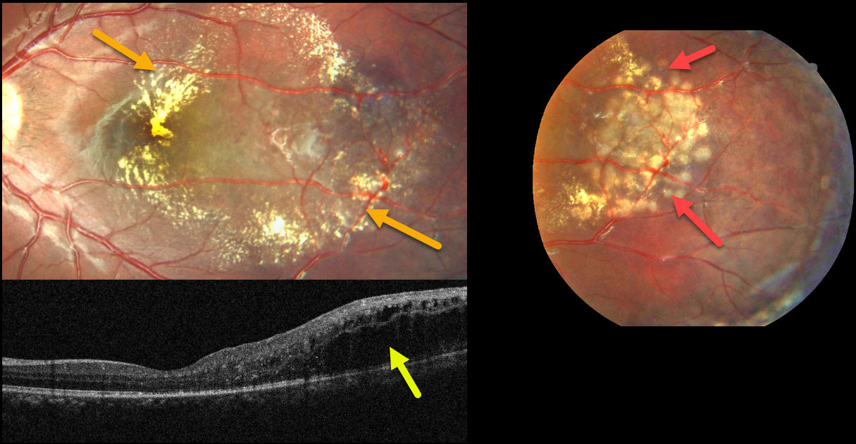
This healthy 8YO boy presented for a routine eye examination. Vision was 20/20 in his normal right eye and 20/30 in his left eye.
Fundus photography shows telangiectatic vessels in the temporal macula associated with surrounding lipid. The lipid is particularly dense centrally.
OCT scanning shows temporal, mostly outer retinal edema. The macular center looks remarkably normal given the fundoscopic appearance. Focal macular laser to the telangiectatic lesions was applied
Learning Points:
Described by George Coats in 1908, Coats disease is an idiopathic disorder typically unilateral in males. It is characterized by retinal vascular telangiectasia involving capillaries, arterioles, and venules.
Patients can present with a wide range of findings, from minimal exudation to total exudative retinal detachment. The leaking vessels are usually treated to prevent exudative complications.
Thermal laser photocoagulation is usually the initial modality, but anti-VEGF injections can also be used to stabilize the vascular leakage. Patients will often require multiple rounds of treatment.
See Sigler et al for a review of the current management (Survey Ophthalmology 2014;59:30-46).



