

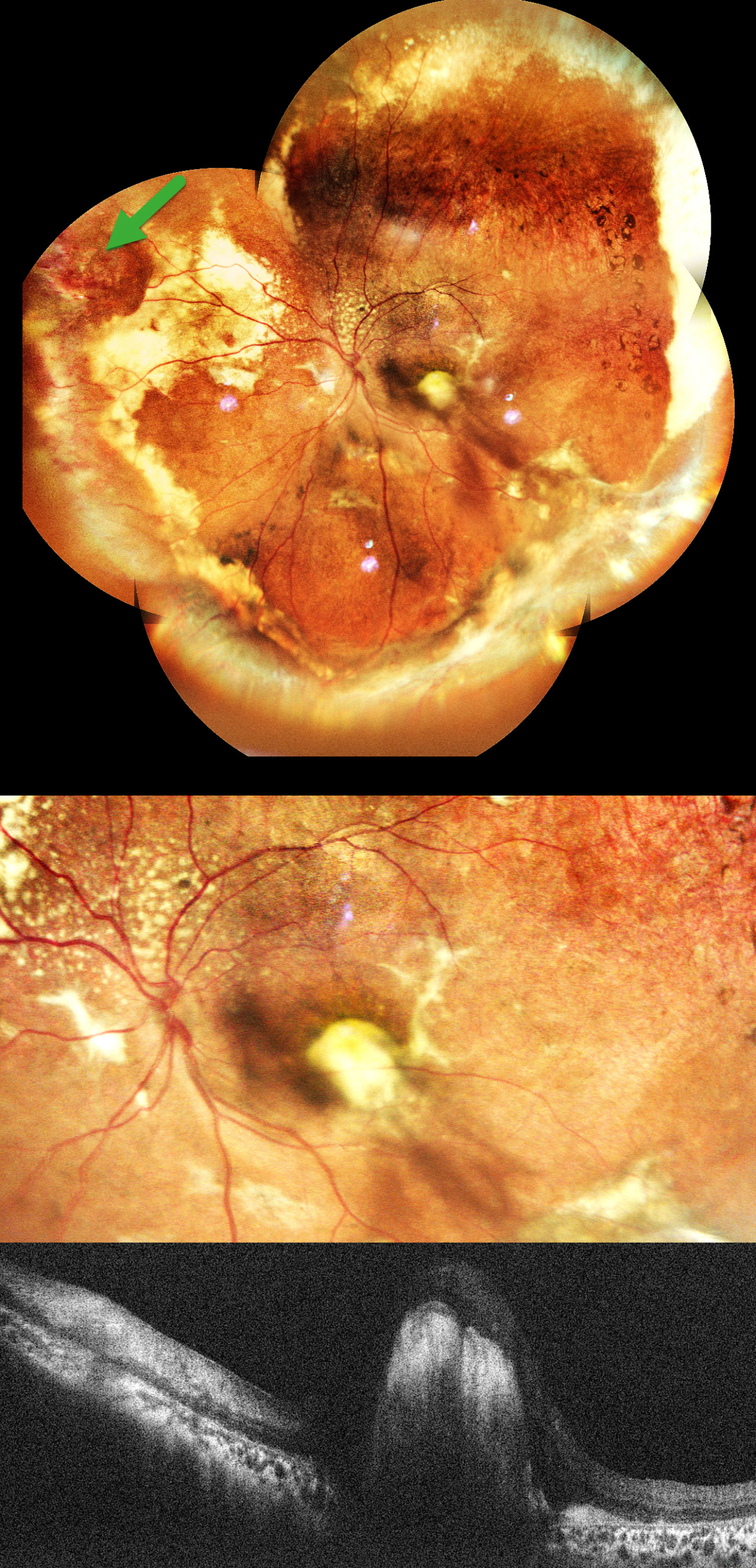
This healthy 11YO boy was brought in by his parents with a 1.5-year history of vision loss and outward deviation. Vision was 20/15 in his normal OD and light perception OS. Xanthocoria was noted on the pupillary light reflex.
Pseudocolor SLO imaging shows extensive peripheral subretinal lipid in all quadrants with telangiectatic and aneurysmal capillary dilations, most prominent in the nasal periphery. A superonasal orange-red elevated vasoproliferative mass is noted in the superonasal midperiphery. Pigmentary changes and RPE atrophy are observed in the superotemporal mid-peripheral retina. There is a large subfoveal lipid plaque that appears as a hyperreflective nodule on OCT. An exudative subtotal retinal detachment involving the inferior and nasal quadrants was noted clinically.
Learning Points:
Coats disease is characterized by vascular changes throughout the retinal vasculature, including the arteries, the capillary bed, and the veins. Larger dilated vessels in Coats can sometimes mimic angiomas, vascular tumors, or exudation from retinoblastoma. The presence of retinal telangiectasia can help establish the diagnosis of Coats in these cases.
Coats is usually unilateral in males. However, recent ultra-widefield angiographic studies indicate subtle vascular changes mostly in the temporal periphery of the fellow eye (Brockmann et al, BJO 2021;105:1444-1453).
Laser photocoagulation can be used to directly treat leaking telangiectatic vessels and ischemic retina. Intravitreal anti-VEGF injections can also be used as an adjunctive treatment. Pars plana vitrectomy (PPV) with external drainage of subretinal fluid plays an important role in the management of advanced Coats disease, particularly in cases with extensive exudative retinal detachment.
In our patient, pars plana vitrectomy with external drainage of subretinal fluid, along with endolaser/cryotherapy to leaking aneurysms, and an intravitreal anti-VEGF injection was advised.



