

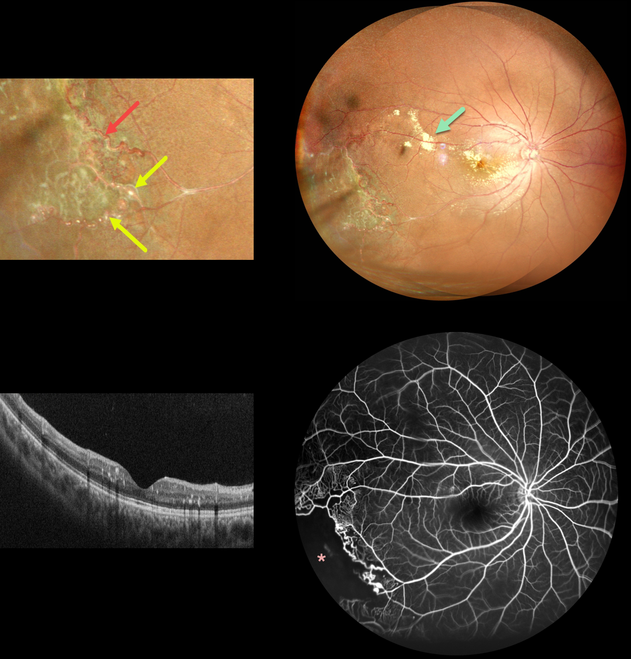
This 11YO boy presented with these classic unilateral findings for Coats disease. An ultrawidefield image of the right eye shows scattered submacular and subretinal lipid posteriorly with distal featureless ischemic retina inferotemporally.
More peripherally, there are both large vessel changes, including beading and multiple macroaneurysms, with small vessel telangiectatic changes.
OCT scanning is remarkably normal except for some scattered specks of hyperreflective intraretinal lipid. Fluorescein angiography shows peripheral temporal telangiectasia with more distal ischemia, along with some subtle superotemporal macular telangiectatic changes.
Sector scatter laser was recommended.



