

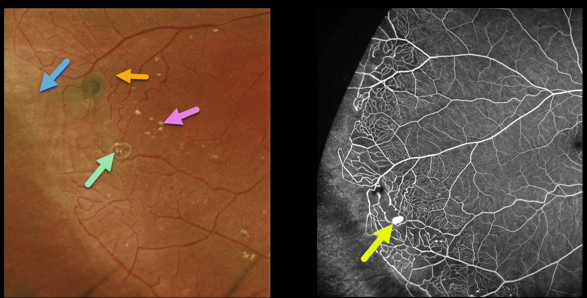
This healthy 13YO boy presented with these asymptomatic findings in his right eye. Vision was 20/20 OU.
Optos imaging shows classic findings for Coats disease in the temporal periphery, including telangiectatic vessels, a macroaneurysm, scattered mild lipid, and ischemic retina more distally. Fluorescein angiography more dramatically illustrates these vascular changes. The macroaneurysm stains.
Learning Points:
Historically considered a unilateral disease, more recent ultra-widefield angiographic studies show subtle vascular changes, mostly in the temporal periphery (Brockmann et al, BJO 2021;105:1444-1453). Our patient’s left eye, however, was completely normal clinically and angiographically.
Due to the lack of significant exudation, we are currently monitoring this patient twice yearly.



