

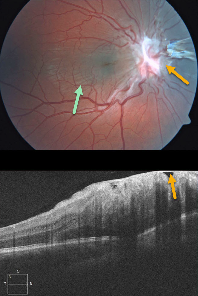
This 7YO boy presented without visual symptoms and 20/50 vision OD. The left eye was normal.
Contracted fibrotic tissue overlying the optic nerve is causing distortion of the surrounding retina, with striae visible through the macula. Some of this fibrosis may also be pigmented nasal to the nerve.
This case represents either a combined hamartoma of the retina and RPE or an unusual pucker centered over the optic nerve.
Learning Points:
Combined hamartomas are classically described as benign congenital lesions composed of glial cells, vascular tissue, and pigmented retinal pigment epithelial cells. However, more recent data indicate that these lesions primarily arise from the inner retinal layers.



