

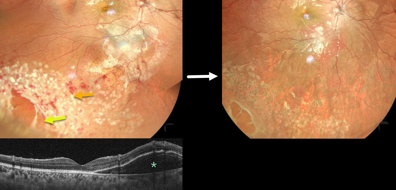
This 13YO male was hit in his right eye with a cricket ball. When he presented later that day vision was 20/30.
Color photography shows a large area of commotio retinae with scattered retinal hemorrhages involving the inferotemporal fundus. There is a poor foveal reflex with possible nasal macular subretinal fluid, which is confirmed on OCT. Several atrophic retinal breaks are noted peripherally. Immediate prophylactic laser was applied around all breaks. Seven weeks later, the commotio and hemorrhages are resolved, and all breaks are well-surrounded with laser scarring. Vision was 20/20.
Although blunt trauma is a common cause of retinal breaks, acute or subsequent dissolution of the retina within areas of commotio with secondary holes is quite rare in our experience (Cox et al Arch Ophthalmol 1966;76:678-685 and Longstaff et al Br J Ophthalmology 1987;71:375-376). The acute white retinal changes usually resolve within several weeks. With resolution, the fundus appearance can normalize. With more severe injury, outer retinal loss can allow RPE cells to migrate into the retina, giving a pseudo-retinitis pigmentosa appearance.
Transient subretinal fluid is an uncommon finding with blunt trauma. Injury to the choroid and RPE allows fluid to accumulate in the subretinal space and outer retina (Mishra et al, Journal of Vitreoretinal Diseases 2021;5:165-169).



