

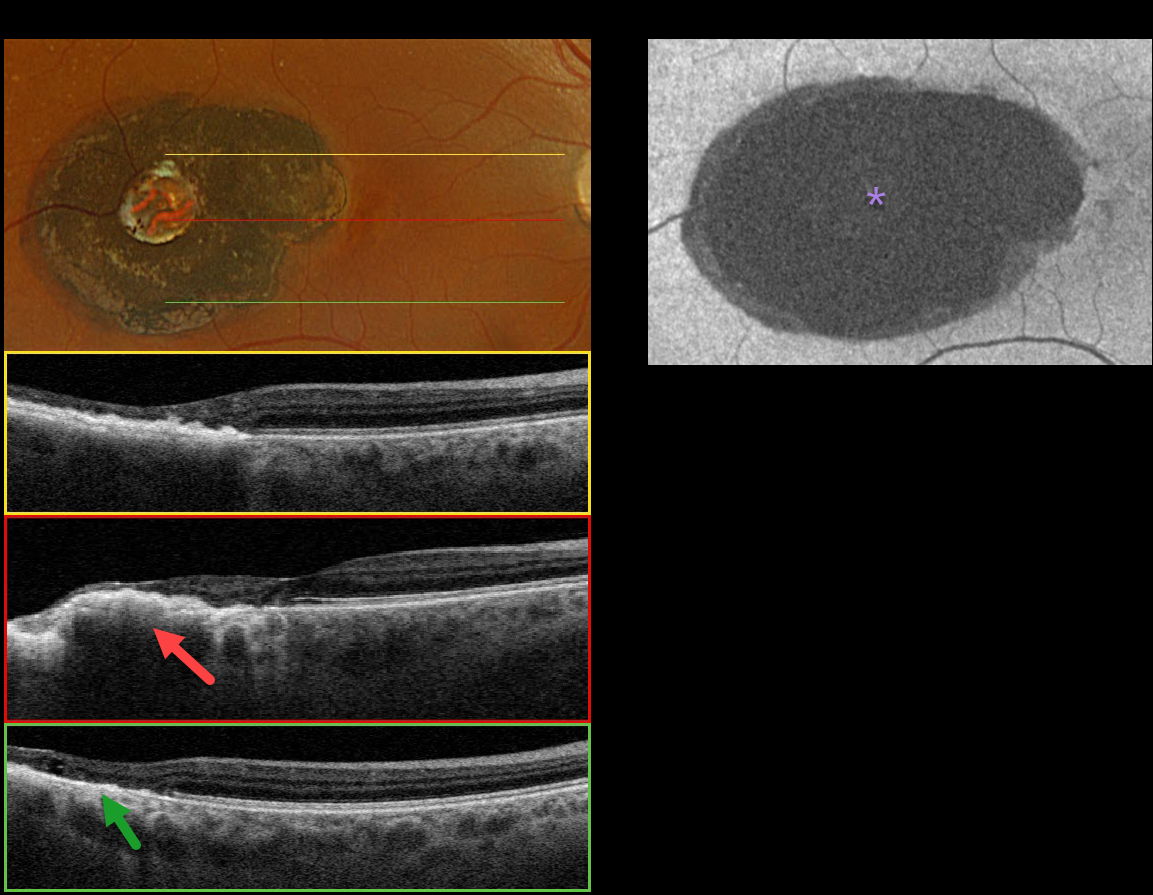
This 57YO female was referred for an asymptomatic pigmented lesion in her right macula. On further questioning, she was told there was something in the back of her eye since childhood due to congenital toxoplasmosis. Vision was 20/20 in her right eye and 20/20 in her left eye.
Optos color RGB imaging shows a well-defined, jet-black lesion in the temporal macula. It has a well-defined hyper/hypopigmented ring along with a central round area of depigmentation.
Triton swept-source OCT shows an irregularly thickened, elevated, and hyperreflective RPE with variable overlying outer retinal atrophy. There is no coloboma. Fundus autofluorescence (FAF) shows uniform hypo-FAF. Observation was recommended.
Learning Points:
Our patient had previously been diagnosed with toxoplasmosis scarring. However, we believe a CHRPE lesion is more likely, given the smooth, well-demarcated hyper/hypopigmented rim typical of CHRPE, the central lacuna, and the lack of colobomatous changes often found in congenital toxoplasmosis lesions (Kumar et al, AJO 2019;200:47-56).
Although CHRPE lesions can enlarge with time (Shields et al, Ophthalmology 2003;110:1968-1973), they carry virtually no malignant potential. Histologic RPE hypertrophy can present with several clinical manifestations.
The typical isolated CHPRE lesions (this case) and bear tracks are not associated with intestinal polyps or cancer, and thus require no systemic evaluation. Only the multiple, small comet-shaped lesions are part of the familial polyposis spectrum.



