

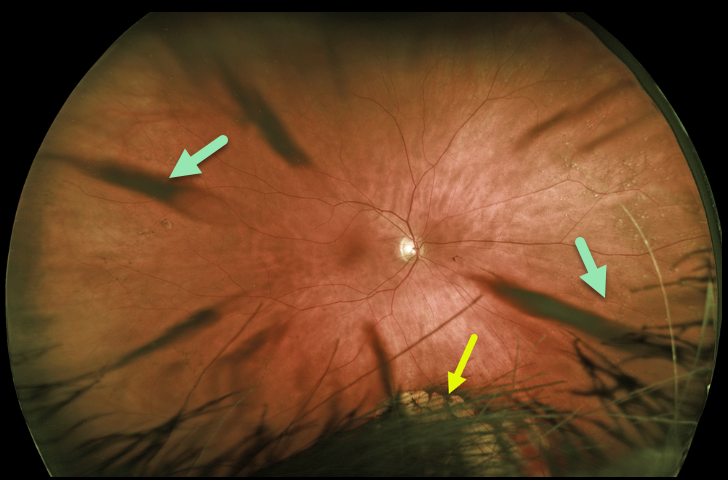
This patient’s Optos image shows beautiful cortical cataract spoke artifacts along with congenital hypertrophy of the RPE (CHRPE) inferiorly, which is mostly obstructed by the patient’s hair and eyelashes.
Learning Points:
Cataracts or other anterior segment findings can create artifacts in fundus photography or optical coherence tomography.
Superior eyelashes often obscure the inferior retinal periphery on Optos imaging, so the technician needs to remind the patient to open their eyes wide and sometimes manually elevate the lid.



