

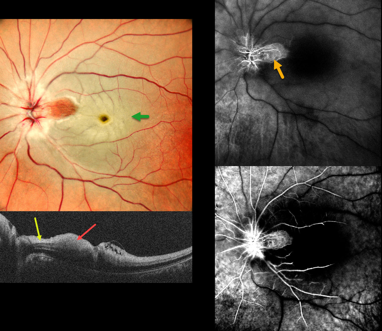
This 37 YO male presented with acute vision loss in his left eye 3 days earlier. Vision was 20/20 in his normal OD and 20/200 in his OS.
Pseudocolor SLO imaging shows a fresh central retinal artery occlusion (CRAO) with sparing of a small area of temporal peripapillary retina supplied from a patent cilioretinal artery. OCT scanning shows opaque, hyperreflective, thickened inner retinal layers, except for a small area of more normal retina within the cilioretinal sparing.
Fluorescein angiography shows profound diffuse retinal nonperfusion and delayed filling except for the nasal macula. Workup elsewhere included an MRI showing left ICA and left MCA chronic infarcts.
Learning Points:
The inner two-thirds of the neurosensory retina is supplied by the central retinal artery, with the choroid supplying the RPE and photoreceptor layer. Acute CRAO, therefore, presents with a white edematous inner retina that spares the fovea. Cilioretinal arteries, which arise from the posterior choroidal circulation, are present in up to 50% of individuals, and bilateral in about 25% (Schneider et al, Acta Ophthalmologica 2021; 99:e310-e318).



