

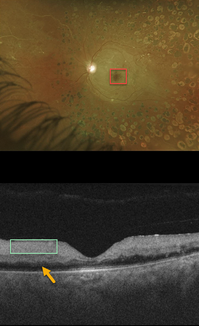
This patient presented with acute vision loss from a fresh central retinal artery occlusion (CRAO). Of note, this patient also has involuted PDR following full PRP.
The cherry red spot is caused by the unaffected foveal retina (receiving intact choroidal blood flow) being surrounded by more distal white and ischemic retina no longer receiving circulation from the central retinal artery.
Although the retina is somewhat thickened on OCT, the acutely ischemic inner two-thirds of the retina, which is supplied by the central retinal artery, is extremely hyperreflective. Less light is available to illuminate the deep retina, which is therefore darker than normal with “pseudo-edema.”
Learning Points:
Patients with acute retinal ischemia (acute CRAO, acute branch retinal artery occlusion or transient monocular vision loss) need to be sent immediately to the nearest stroke center due to the risk of pre-existing recent asymptomatic stroke or impending stroke (see Biousse et al, Ophthalmology 2018;125;1597-1607 or our post https://odsonfb.com/amaurosis-brao-and-crao-the-medical-emergency-many-eye-doctors-still-dont-know-about/).



