

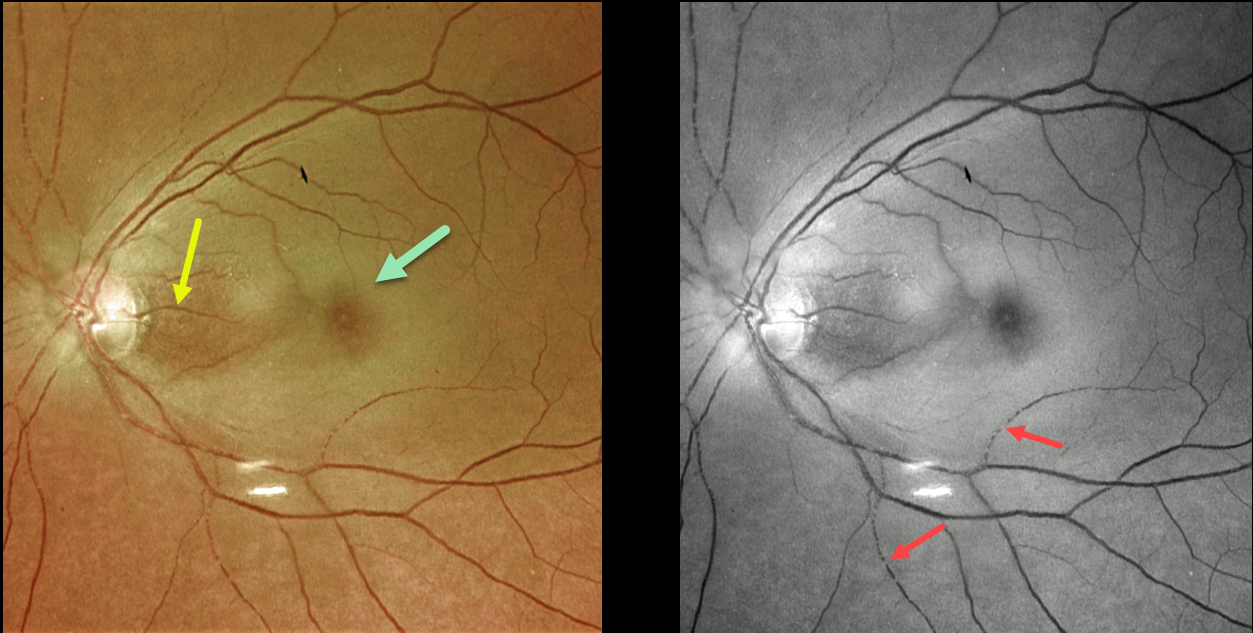
This 73YO female presented with counting-fingers vision secondary to an acute central retinal artery occlusion (CRAO).
The foveal retina is still perfused by the underlying choroid, resulting in the classic cherry red spot appearance. The nasal macula is also preserved due to the presence of a cilioretinal artery, which unfortunately does not supply the macular center. The Optos green channel best images the stagnant blood column.
Our patient’s MRI revealed several acute diffuse embolic strokes. Carotid duplex ultrasound showed severe bilateral carotid stenosis. In addition, her blood glucose was 357, and her erythrocyte sedimentation rate was 94. She was also started on systemic prednisone by the ER until giant cell arteritis could more definitively be ruled out.
Learning Points:
Patients with acute retinal ischemia (defined as transient monocular vision loss, acute BRAO, or acute CRAO) need to be emergently referred to a stroke center.
This is especially urgent with an acute CRAO since about 75% of patients have already developed a recent stroke.



