

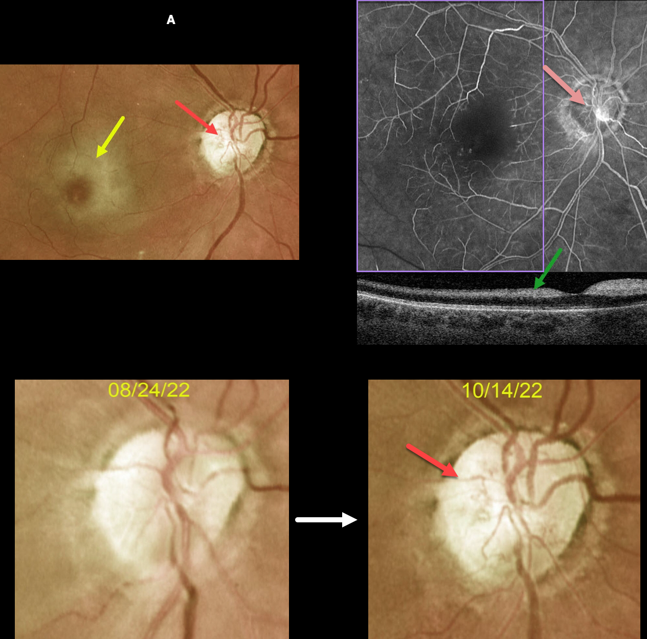
This 63YOF initially presented with an acute central retinal artery occlusion in her right eye. Emergency stroke referral revealed two prior strokes with a new asymptomatic stroke.
She returned two months later. There was subjective visual improvement, although vision remained at hand motion. Although the retinal opacification in general was improving, the perifoveal retina was more opaque with a pronounced cherry red spot.
New fine vessels were noted on the nerve. These vessels did not leak on fundus fluorescein angiography. Marked angiographic ischemia is also noted. OCT scanning shows increased reflectivity from the ischemic inner retina.
Learning Points:
Collateral vessels are dilated, pre-existing vessels that bypass an occlusion. These are seen in the posterior pole most commonly following retinal venous occlusions and are exceedingly rare with arterial occlusions.
The endothelial tight junctions are intact, so they don’t leak angiographically. This helps differentiate them from neovascularization, which does leak.



