

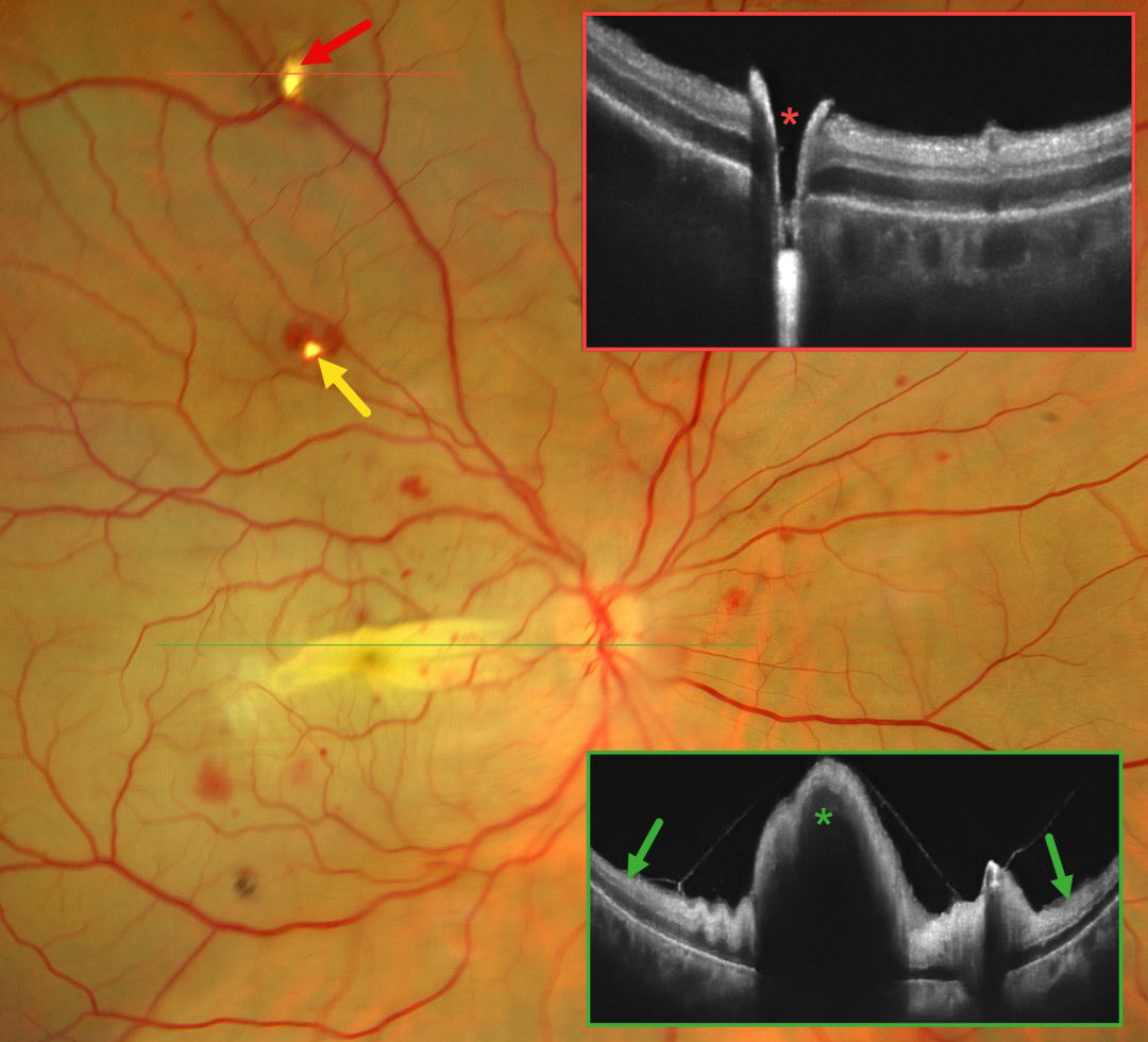
This 42YO female immediately lost vision in her right eye 4 days earlier following pterygium excision surgery performed under peribulbar anesthesia elsewhere. Vision in our office was light perception.
Fundus photography shows an opaque retina throughout the posterior pole consistent with an acute central retinal artery occlusion (CRAO). There is a horizontal retinal fold throughout the macula with scattered blot retinal hemorrhages. Entry and exit needle penetration sites are noted. OCT scan through the superior entry site confirms a full-thickness retinal break. Macular OCT shows diffuse inner retinal hyperreflectivity with elevation through the fold.
Learning Points:
Peribulbar anesthesia is considered to have a lower risk of inadvertent globe perforation, although this risk is not zero (Lim et al., J Clin Ophthalmol 2021;5:414-416). CRAO after such an injury is exceedingly rare and was reported by Lake et al. (J Cataract Refract Surg 2003;29:2234-2235), who felt the occlusion might have resulted from increased IOP due to intraocular anesthetic. A CRAO was also reported by Gyasi et al. following peribulbar anesthesia for pterygium excision, although they found no evidence of globe perforation (Ghana Medical Journal 2012;46:48-50).



