

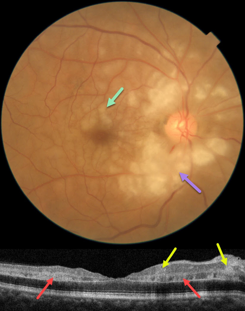
This 60YO male presented with an abrupt, painless loss of vision in his right eye 1 week earlier. There was no prior ocular history or recent history of trauma. Vision was 20/30 in his right eye, and 20/20 in his normal left eye.
Fundus photography shows patches of mostly inner-peripapillary ischemia, with deeper ischemia throughout the macula. This is shown on OCT as patches of inner and mid-retinal hyperreflectivity. Fluorescein angiography showed a markedly delayed transit time (not shown).
We immediately referred him to his cardiologist for an emergency stroke protocol evaluation.
Learning Points:
Purtscher retinopathy was first described by Otmar Purtscher in 1910. Although originally described in a man who fell from a tree with cranial trauma, these white patches of retinal ischemia and hemorrhages are found in numerous other etiologies, including pancreatitis, chest trauma, and collagen vascular diseases.
The findings are called Purtscher retinopathy when due to trauma, and Purtscher-like retinopathy when seen from other causes.
Our patient’s fundus appearance most resembles Purtscher-like retinopathy, with patches of inner- and middle-retinal ischemia. However, given the acute symptoms and delayed angiographic filling time, we believe our patient most likely has an incomplete central retinal artery occlusion (CRAO).
See Mangla et al for the OCT findings in CRAO of varying severities (Int J Retina Vitreous 2023;9;37).



