

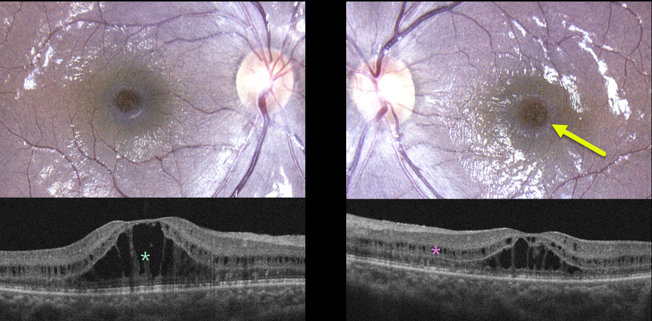
This healthy 12YO girl was referred for bilateral macular changes. She was having some mild subjective central vision loss. Vision was 20/40 OD and 20/30 OS. There was no ocular family history.
Color photography shows symmetrical radiating foveoschisis. OCT scanning shows central outer nuclear layer schisis with milder, more peripheral inner nuclear layer schisis.
Learning Points:
X-linked retinoschisis is caused by a mutation of the RS1 gene, which is located on the X chromosome and encodes for retinoschisin, a protein likely involved in retinal cellular adhesion (Heymann et al, Progress Retinal Eye Research 2023;95;101147).
Although usually transmitted as an X-linked recessive disorder found in males, non-sex-linked foveoschisis is present in other heritable disorders, such as enhanced S-cone syndrome and autosomal recessive bestrophinopathy, or non-heritable disorders, such as stellate nonhereditary idiopathic foveomacular retinoschisis.
Genetic testing of our patient revealed two pathogenic mutations of CRB1. Although few cases have been described (Vincent et al, Invest Ophthalmol Vis Sci 2016;57;2637-2646), CRB1-related foveoschisis appears to have a high female preponderance.
Similar to X-linked retinoschisis, these patients tend to maintain good vision into early adulthood, but can progress to secondary macular atrophy later in life with collapse of the schisis.
Thankfully, the foveoschisis phenotype represents the mildest of CRB1-related ocular diseases, which include Leber’s congenital amaurosis, retinitis pigmentosa, early-onset retinal dystrophy, and cone-rod dystrophy (Varela et al., AJO 2023;246:107-121).



