

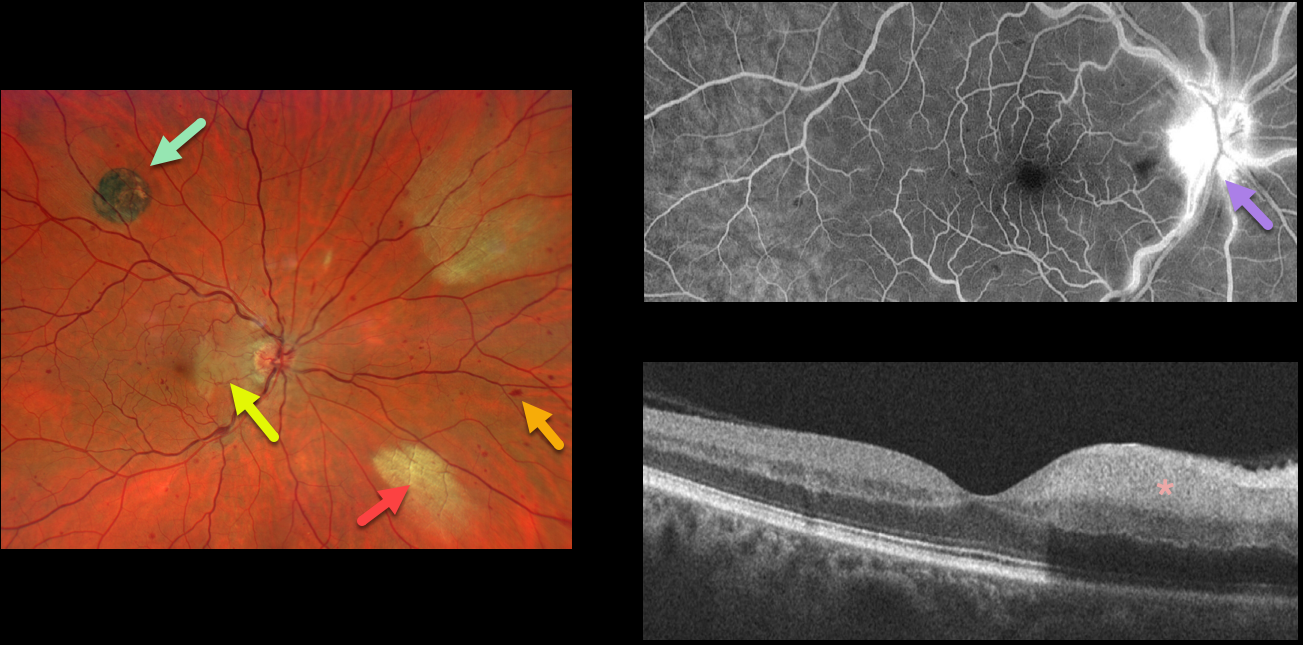
This 52YO male presented with acute visual symptoms in his right eye. Vision was 20/20.
Optos imaging shows a non-ischemic central retinal vein occlusion (CRVO) with mild scattered retinal hemorrhages in all quadrants. An acute nasal macular branch retinal artery occlusion (BRAO) is noted, along with a small round patch of congenital hypertrophy of the RPE (CHRPE) superotemporally, and two patches of myelinated nerve fiber layer (NFL) nasally.
Fluorescein angiography shows a well-perfused central retinal vein occlusion (CRVO) with late optic nerve leakage. OCT scanning shows hyperreflectivity of the inner nasal retina.



