

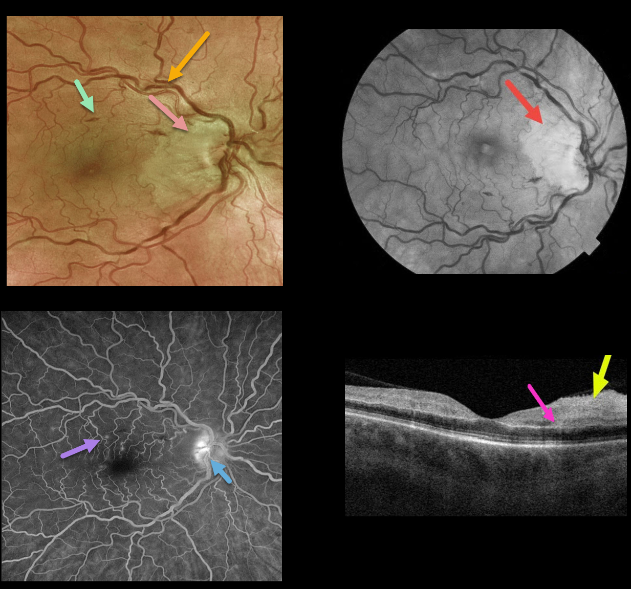
This 50YO female presented with counting finger vision in her right eye due to an acute central retinal vein occlusion (CRVO) with a nasal macular retinal artery occlusion (RAO).
Optos fundus photography shows retinal vascular tortuosity, mild retinal hemorrhages, a mildly swollen nerve, and white ischemic nasal macular retina. The ischemic retinal changes are most apparent on the red-free photo.
Fluorescein angiography (FA) shows some mild focal areas of macular leakage with late staining of the temporal disc.
OCT shows areas of inner retinal edema and hyperreflectivity. There is also a small area of mid-retinal hyperreflectivity consistent with paracentral acute middle maculopathy (PAMM).
Learning Points:
Cilioretinal artery occlusion is usually non-embolic and seen in association with giant cell arteritis, secondary to a CRVO, or as an isolated event. The occlusion may be due in part to arterial compression from a swollen nerve.
Pichi et al felt that CRVO PAMM-type lesions, as seen in our patient, are due to hypoperfusion from increased downstream intraluminal pressure (Br J Ophthalmol 2019;103:1137-1145).
We couldn’t find a definite cilioretinal artery in our patient, although the ischemia was within the distribution of one if one were present. Interestingly, the nasal macular capillaries appeared perfused on FA. However, FA captures just the inner retinal capillary plexus.
Although the OCT shows evidence of acute inner capillary plexus ischemia, the angiographically perfused inner retina supports a more PAMM-type mechanism in our case.



