

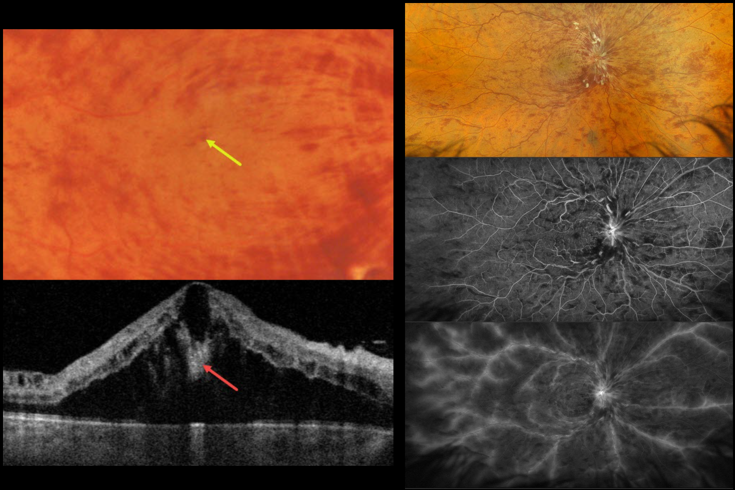
This healthy 52YO male presented with recent severe vision loss in his right eye. Vision was 20/400 OD and 20/20 in his normal OS.
Color photography and Optos RGB imaging show a central retinal vein occlusion (CRVO) with marked retinal hemorrhages and macular edema. Inner blood-filled retinal cysts are noted on funduscopy and swept-source OCT. On fluorescein angiography, the occlusion is fairly well perfused with late leakage from the nerve, macula, and retinal veins. Monthly anti-VEGF therapy was started.
Learning Points:
Cicinelli et al recently reported that central bouquet hemorrhages in retinal vein occlusion are associated with poor visual outcomes due to localized effects of elevated venous pressure and macular ischemia (IOVS 2025;66(12):62).
Prior to the introduction of intravitreal injections in the early 2000s, there were essentially no treatments for CRVO-related macular edema. Anti-VEGF injections are now generally the initial treatment of choice, with about 50-60% of eyes gaining at least 3 lines of vision. Intravitreal steroids, usually Ozurdex, are more often used as secondary treatment. Macular grid laser photocoagulation is not performed because it has been shown to confer no visual benefit in the Central Vein Occlusion Study Group (Ophthalmology 1995;102:1425-1433).



