

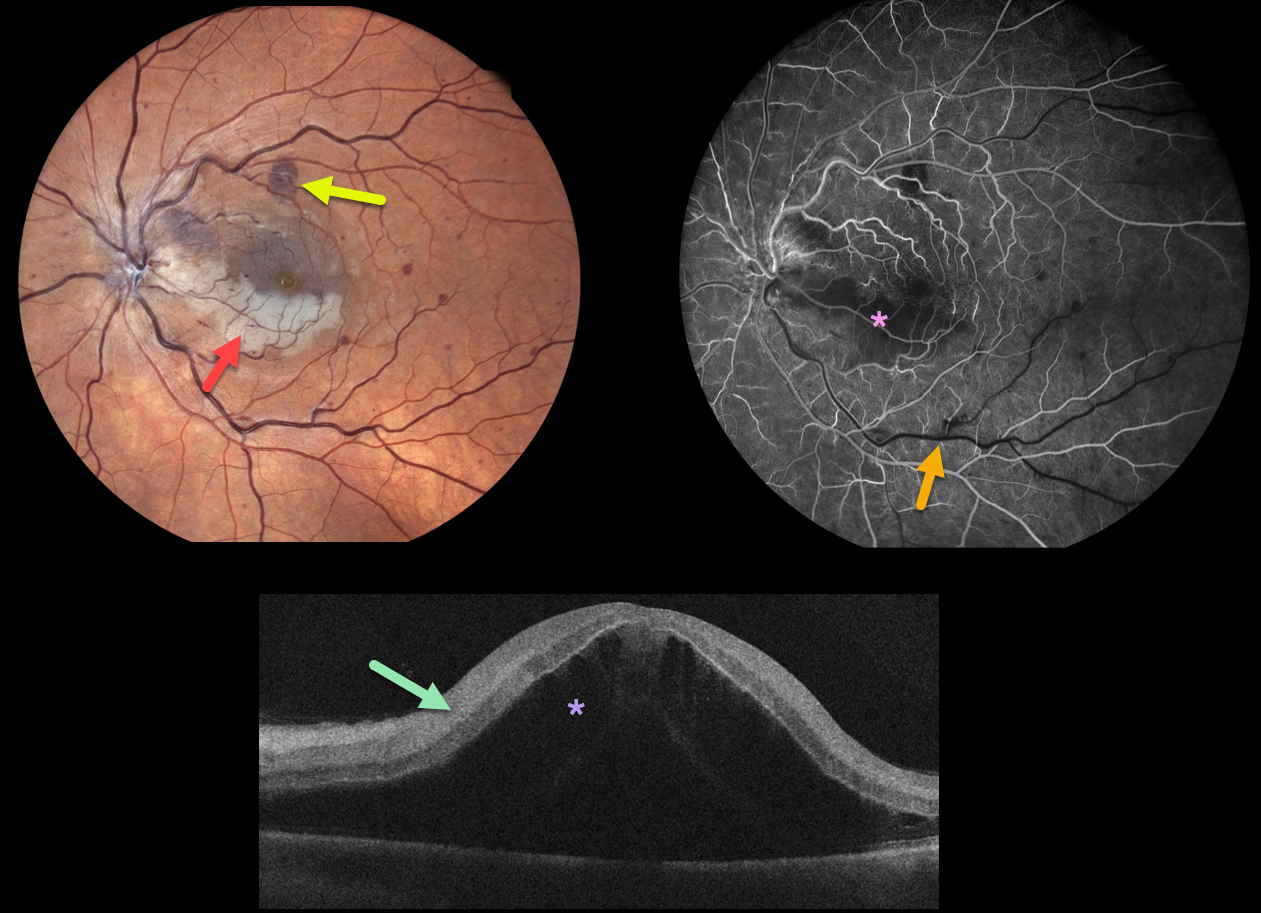
This 49YO male woke with counting finger vision in his left eye. Fundus photography shows a fresh central retinal vein occlusion (CRVO) with mild retinal hemorrhages. An acute inferior macular branch retinal arterial occlusion (BRAO) is also noted.
OCT B-scan shows marked inner retinal hyperreflectivity and severe macula edema.
Fluorescein angiography in the venous laminar phase shows inferior macular hypofluorescence from combined blockage from the ischemic white retina, along with a filling defect from the BRAO. The retinal perfusion elsewhere is good.
Some patients with CRVO will develop a secondary non-embolic BRAO caused by compression from optic nerve edema. However, our patient’s optic nerve did not appear that swollen. Anti-VEGF therapy was started for the severe macular edema.



