

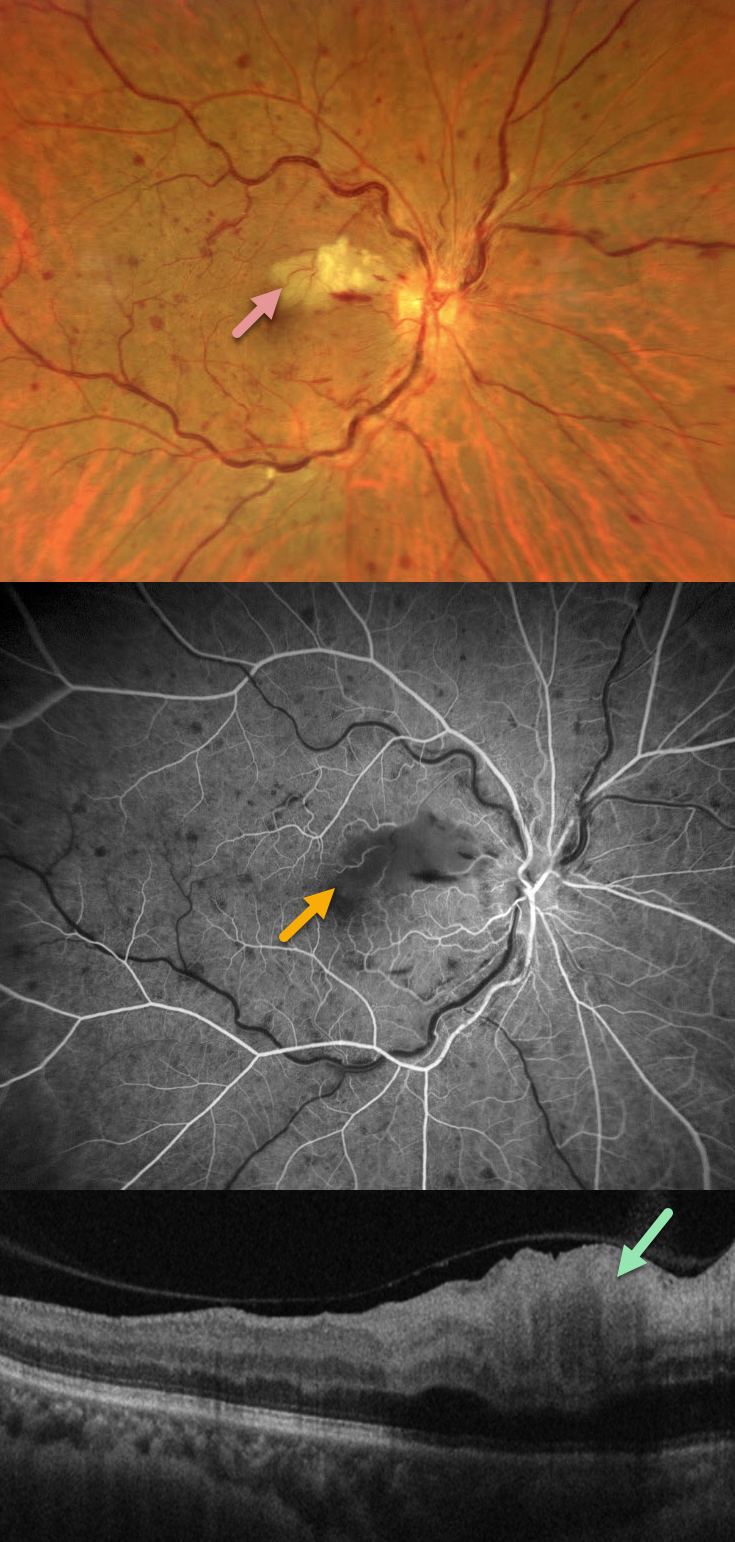
This 58YO female with a history of hypertension and diabetes presented with sudden painless vision loss in her right eye. Vision was 20/25 OD and 20/20 in her normal OS.
Fundus photography shows a non-ischemic central retinal vein occlusion (CRVO) with dilated tortuous veins, mild scattered retinal hemorrhages, and a superonasal macular branch retinal artery occlusion (BRAO). The BRAO shows inner retinal hyperreflectivity on OCT. Fluorescein angiography shows good perfusion throughout the distribution of the CRVO, with a filling defect from the BRAO.
Learning Points:
Some patients with CRVO will develop a secondary non-embolic cilioretinal artery occlusion caused by either compression from optic nerve edema or from a sudden rise in intraluminal retinal capillary bed pressure (Hayreh et al, Retina 2008;28;581-594).



